EP0103477A2 - Method of transferring tissue section from support to microscope slide - Google Patents
Method of transferring tissue section from support to microscope slide Download PDFInfo
- Publication number
- EP0103477A2 EP0103477A2 EP83305306A EP83305306A EP0103477A2 EP 0103477 A2 EP0103477 A2 EP 0103477A2 EP 83305306 A EP83305306 A EP 83305306A EP 83305306 A EP83305306 A EP 83305306A EP 0103477 A2 EP0103477 A2 EP 0103477A2
- Authority
- EP
- European Patent Office
- Prior art keywords
- layer
- tissue section
- substrate
- process according
- tissue
- Prior art date
- Legal status (The legal status is an assumption and is not a legal conclusion. Google has not performed a legal analysis and makes no representation as to the accuracy of the status listed.)
- Granted
Links
Images


Classifications
-
- G—PHYSICS
- G01—MEASURING; TESTING
- G01N—INVESTIGATING OR ANALYSING MATERIALS BY DETERMINING THEIR CHEMICAL OR PHYSICAL PROPERTIES
- G01N1/00—Sampling; Preparing specimens for investigation
- G01N1/28—Preparing specimens for investigation including physical details of (bio-)chemical methods covered elsewhere, e.g. G01N33/50, C12Q
Definitions
- This invention relates to a method for transferring thin sections of tissue specimens, prior to staining and mounting processes, from a support to a microscope slide.
- Very thin slices of animal and plant tissues are prepared for many different kinds of microscopic studies by sectioning using various kinds of microtomes. While the tissue may be cut fresh, the soft and compliant nature of most fresh tissue makes the cutting of undistorted thin sections very difficult. Often, the tissue is cut on a freezing microtome or in a cryostat at temperatures below 0°C. (32 0 F.,), the hardness of the frozen water within the tissue allowing sections as thin as a few micrometers to be cut relatively easily. As these frozen sections are brittle and friable, they are difficult to handle and process further. To simplify sectioning of tissue, a number of procedures have been developed which produce a block of supported tissue which has superior sectioning properties and produces high quality, relatively easy-to- handle tissue sections. Such procedures typically involve:
- a process for transferring a thin section of a tissue specimen, supported on a substrate by a first pressure-sensitive adhesive layer, from said substrate and mounting it on a microscope slide having at least a portion of one surface coated with a second pressure sensitive adhesive layer, said second layer being polymerisable comprising the steps of:
- a tissue specimen supported on, for example, a flexible piece of pressure-sensitive adhesive tape is brought into contact with a microscope slide over whose contacting surface a film of pressure-sensitive adhesive, capable of further polymerisation, has been layered.
- Techniques by which flexible tape can be applied to such specimens to aid sectioning have been described, for example, in the above- identified publications by Palmgren, Beckel, Gowers et al and Wedeen et al, respectively.
- the layer is then polymerised, usually within about 1 to 3 minutes, and then the tape may, for example, be submerged in a solvent which weakens the supporting tape backing and/or supporting-tape adhesive-layer which are washed or peeled away, to fully expose the tissue specimen.
- the polymerisable layer and its bonds to the section have sufficient strength after curing to allow peeling of the insoluble tape backing without damaging the section and to thereafter firmly support the specimen on the microscope slide.
- the polymerisable layer preferably has only curable components whose respective diffusion coefficients, before and during cure, are sufficiently low that the diffusion thereof into the specimen during the curing process is minimal, and/or a viscosity sufficiently large that flow into the specimen on application of finger-pressure is minimal, that is less than 1 micrometer in 30 minutes.
- the layer forms a positive bond between the microscope slide and specimen section, to permit ease of handling during subsequent processing of the supported section.
- the substantially entire interior structure of the tissue section remains exposable to processing.
- the polymerised bonding layer is not affected, in any way, by such processing.
- the polymerised layer preferably has a refractive index matched to the refractive index of the unstained tissue, e.g., from about 1.53 to 1.57, and to the mounting medium conventionally used to envelop and to attach the coverslip over the specimen at the completion of the processing.
- the specimen is stained during processing to highlight particular components thereof.
- the respective refractive indices of the various components of the completed microscope slide are substantially the same, there is no optical interference introduced to degrade the image in the subsequent microscopic examination of the tissue.
- the present invention provides a method for positively attaching a tissue to a microscope slide by a pressure-sensitive polymerisable layer, the curable components of such layer prior to and during polymerisation penetrating only very minimally into such specimen and, when polymerised, having a compatible index of refraction and neither affecting'nor being affected by any subsequent chemical or physical treatment of the supported tissue.
- a thin tissue section 9 surrounded and infiltrated with paraffin 10 is supported on and adhered to the surface of a flexible tape or substrate 12, by a pressure-sensitive adhesive layer 14.
- Section 9 is located within a window or opening in a spacer 13 adhered to layer 14.
- spacer 13 facilitates the cutting of a thin section 9 by a microtome.
- a microscope slide 16 which supports on its upper surface a pressure-sensitive adhesive layer 18.
- Layer 18 can be formed of, for example, a heat-curable polymerisable material or a photopolymerisable material.
- layer 18 is photopolymerisable, e.g. by exposure to U.V. light of wavelengths in the range of 320 nm-390 nm.
- Figure 2 further illustrates the inversion of tape 12 so as to juxtapose specimen 9 and layer 18.
- Specimen9 and layer 18 are subsequently contacted.
- a slight pressure is applied, e.g. manually.
- such pressure can be applied progressively along the specimen 9, to ensure a smooth contact thereof with layer 18.
- Layer 18 preferably is formed to have the characteristics of:
- the laminate 20 is then immersed is a solvent solution 22, such as xylene, contained in beaker 24.
- a solvent solution 22 such as xylene
- Such immersion serves to rapidly soften-the layer 14 on tape 12 and allows for a gentle manual peeling of tape 12 from over specimen 9 while laminate 20 is immersed in beaker 24.
- Exposed layer 14 and paraffin 10 dissolve as, or within on minute after, tape 12 is peeled away.
- the specimen section 9 is now fully exposed.
- the solvent solution 22 may be selected, so as to be effective to dissolve both substrate 12 and adhesive layer 14, so as to avoid the need of peeling substrate 12, as described.
- the layer 18 on slide 16 consists of a mixture of very high molecular weight reactive oligomers (typically, each with two terminal acryl or methacryl groups).
- the high molecular weight ensures a high viscosity and a low diffusion coefficient. If the molecular weight is too high, there will be insufficient tack.
- the acryl or methacryl groups permit radical-initiated polymerisation to crosslink the oligomers into a highly insoluble polymer.
- the mixture is composed of one oligomer with a cured refractive index of greater than 1.560 (e.g. an epoxy-acrylate oligomer with many bisphenol A or other high aromatic residues to raise the refractive index) and another with a cured refractive index less than 1.530 (e.g. a urethane acrylate oligomer).
- cured resins must be extremely resistant to xylene, alcohol and water. By mixing them in appropriate proportions, any desired cured refractive index, between 1.530 and 1.560, can be imparted to layer 18, when polymerised.
- the mounting medium for encapsulating specimen section 9 should preferably have a refractive index near 1.55, and layer 18, when cured, a refractive index of between 1.545 and 1.555.
- specimen section 9 can be processed for storing and permanent encapsulation, according to conventional histological techniques.
- specimen section 9 is securely and permanently bound to slide 16 by now-polymerised layer 18, which neither affects nor is affected by exposure to such techniques.
- encapsulation process may be manually effected, for example, as fully described in McManus and Mowry above.
- a number of tissue specimen sections 9, each supported on an individual slide 16 may be processed in automated fashion, for example, as clearly shown and described in an AUTOTECHNICON system as marketed by Technicon Instruments Corporation, of Tarrytown, New York, and described in Technical Publication No. TA 1-0225-10, June 1977, pages 3-10-3-13.
- Figure 4 illustrates a completely mounted specimen section, ready for microscopic viewing.
- Figure 4A illustrates a specimen section 9 mounted by conventional techniques; such specimen is covered with a mounting medium 26 and a coverslip 28 is located on-such medium and over such specimen.
- Figure 4B shows the envelopment of a specimen 9 in a U.V. curable mounting medium 27, as results from the technique disclosed in U.S. Patent 4,120,991.
- a coverslip is not provided. Rather the upper surface of mounting medium 27 is formed so as to be optically flat.
- the adhesive tape used to capture the sections must at least have an adhesive layer which is soluble in a solvent, such as xylene.
- a solvent such as xylene.
- a silicone rubber adhesive as used by Wedeen and Jernow referenced above is satisfactory for frozen sections, and an adhesive tape, like Permacel No. 925 is satisfactory for paraffin sectioning.
Abstract
Description
- This invention relates to a method for transferring thin sections of tissue specimens, prior to staining and mounting processes, from a support to a microscope slide.
- Very thin slices of animal and plant tissues are prepared for many different kinds of microscopic studies by sectioning using various kinds of microtomes. While the tissue may be cut fresh, the soft and compliant nature of most fresh tissue makes the cutting of undistorted thin sections very difficult. Often, the tissue is cut on a freezing microtome or in a cryostat at temperatures below 0°C. (320F.,), the hardness of the frozen water within the tissue allowing sections as thin as a few micrometers to be cut relatively easily. As these frozen sections are brittle and friable, they are difficult to handle and process further. To simplify sectioning of tissue, a number of procedures have been developed which produce a block of supported tissue which has superior sectioning properties and produces high quality, relatively easy-to- handle tissue sections. Such procedures typically involve:
- (1) fixation of the tissue in a solution which insolubilises and hardens the natural polymers of which tissue cells are composed; (2) dehydration of the tissue through a series of water-miscible (e.g. an alcohol) and then paraffin or plastic-monomer-miscible (e.g. toluene or xylene) solvents; (3) infiltration of the tissue by melted paraffin or monomer solution; and (4) embedding by freezing the paraffin or polymerising the monomer to form a solid polymer. Reference is made to Staining Methods, J.F.A. McManus and R. W. Mowry (P.B. Hoeber, Inc., N.Y. 1960) and to Techniques for Electron Microscopy, D. Kay, Ed. (Blackwell Sci. Publ., Oxford, England, 1965) pp. 166-212.
- However, there are certain specimens which remain difficult to section. As the section is cut from the tissue, parts of the cut section tend to fragment and fall from the cut section, or fall from the section as it is removed from the microtome. Reference is made to the following which describe procedures for facilitating the cutting and handling of such difficult-to-cut tissue sections:
- 1. A. Palmgren, Nature, Vol. 174, p. 46, (1954) describes the use of a pressure-sensitive adhesive tape as a sectioning aid for the cutting of very large, hard or brittle specimens. A piece of adhesive tape is applied to the surface of a specimen, either frozen or embedded in a paraffin block, supported in a microtome. Thus, the section, when cut, is supported by the applied tape. The quality of the uncompressed section of hard, brittle and friable tissue thus produced can be far superior to that of a conventional section of the same block of tissue. However, following Palmgren, processing such a section while it remains on the tape, or transferring it to a glass slide (to permit it to be processed thereafter in conventional fashion) involves elaborate, time-consuming and inconvenient methods which can also damage the section.
- 2. W. E. Beckel, Nature, Vol. 184, p. 1584 (1959) describes the use of Scotch brand No. 810 cellulose- acetate-backed adhesive tape in the process described by Palmgren above. The tape-mounted sections are applied, section-side down, on wet conventionally albuminised glass slides. After thorough drying, which requires at least a few hours, the adhesive backing, the adhesive layer and the paraffin are all dissolved in tetrahydrofuran for 30 minutes, leaving only the section adhering to the glass slide and available for further processing by conventional techniques. Alternatively, chloroform for 2 mi-nutes,, followed by xylene for 30 minutes, can be used for dissolving the adhesive backing, the adhesive layer and the paraffin. Beckel also describes a "more rapid method" which uses a film of albumin and a solution of 2 per cent celloidin in methyl benzoate or ethyl alcohol to "cement" the section to the glass slide, followed by 1 minute in chloroform and 10 minutes in xylene to complete the treatment.
- 3. D. S. Gowers and R. E. Miller Nature, Vol. 190, p. 425 (1961) attempted to repeat Beckel's method but found that, with available Scotch brand No. 810 tape, the adhesive could not be dissolved, and with the best alternate available tape, Tuck brand No. 200, safe removal of the tape without damaging the section in solvent took from 1 to 10 hours.
- 4. R. P. Wedeen and H. I. Jernow Am. J. Physiol., Vol. 214, p. 776, (1968) used cyanoacrylate (Eastman 910 "superglue") to attach adhesive- tape-supported frozen sections to radioauto- graphic (photographic) plates. The cyanoacrylate is initially liquid, but polymerises to a solid when pressed into a thin film. As the cyanoacrylate polymer is soluble in xylene and other processing solvents which would cause the section to float free, it is not useful for conventional staining procedures.
- We have now devised a method of transferring thin tissue sections from supports such as adhesive tape, on to microscope slides, by which certain of the disadvantages of prior art procedures are reduced or overcome.
- According to the present invention, there is provided a process for transferring a thin section of a tissue specimen, supported on a substrate by a first pressure-sensitive adhesive layer, from said substrate and mounting it on a microscope slide having at least a portion of one surface coated with a second pressure sensitive adhesive layer, said second layer being polymerisable, comprising the steps of:
- bringing said supported tissue section into contact with said second layer to form a laminate comprising said substrate, said first layer, said tissue section, said second layer, and said microscope slide;
- polymerising said second layer;
- immersing said laminate in a solution in which said first layer is soluble, to facilitate removal of said substrate and said first layer from over said tissue section; and
- subsequently removing said substrate and said first layer to expose said tissue section mounted on said microscope slide.
- In the method of the present invention, a tissue specimen supported on, for example, a flexible piece of pressure-sensitive adhesive tape, is brought into contact with a microscope slide over whose contacting surface a film of pressure-sensitive adhesive, capable of further polymerisation, has been layered. Techniques by which flexible tape can be applied to such specimens to aid sectioning have been described, for example, in the above- identified publications by Palmgren, Beckel, Gowers et al and Wedeen et al, respectively. After the specimen section has been contacted with the polymerisable layer, the layer is then polymerised, usually within about 1 to 3 minutes, and then the tape may, for example, be submerged in a solvent which weakens the supporting tape backing and/or supporting-tape adhesive-layer which are washed or peeled away, to fully expose the tissue specimen. The polymerisable layer and its bonds to the section have sufficient strength after curing to allow peeling of the insoluble tape backing without damaging the section and to thereafter firmly support the specimen on the microscope slide.
- The polymerisable layer preferably has only curable components whose respective diffusion coefficients, before and during cure, are sufficiently low that the diffusion thereof into the specimen during the curing process is minimal, and/or a viscosity sufficiently large that flow into the specimen on application of finger-pressure is minimal, that is less than 1 micrometer in 30 minutes. However, when cured, the layer forms a positive bond between the microscope slide and specimen section, to permit ease of handling during subsequent processing of the supported section. As diffusion and flow into the section is minimal, the substantially entire interior structure of the tissue section remains exposable to processing. Also, the polymerised bonding layer is not affected, in any way, by such processing.
- The polymerised layer preferably has a refractive index matched to the refractive index of the unstained tissue, e.g., from about 1.53 to 1.57, and to the mounting medium conventionally used to envelop and to attach the coverslip over the specimen at the completion of the processing. Conventionally, the specimen is stained during processing to highlight particular components thereof. As the respective refractive indices of the various components of the completed microscope slide are substantially the same, there is no optical interference introduced to degrade the image in the subsequent microscopic examination of the tissue.
- Thus, in a preferred embodiment, the present invention provides a method for positively attaching a tissue to a microscope slide by a pressure-sensitive polymerisable layer, the curable components of such layer prior to and during polymerisation penetrating only very minimally into such specimen and, when polymerised, having a compatible index of refraction and neither affecting'nor being affected by any subsequent chemical or physical treatment of the supported tissue.
- In order that the invention may be more fully understood, reference is made to the accompanying drawings, wherein:
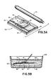
- FIGURE 1 is an isometric view of a thin tissue section supported on the surface of a flexible tape or substrate;
- FIGURE 2 is an isometric view of the substrate of Figure 1 juxtaposed with a microscope slide;
- FIGURE 3 is an isometric view illustrating the super-positioning of the substrate over the microscope slide, so as to laminate the thin tissue section therebetween;
- FIGURE 3B illustrates the removing of the substrate from over the microscope slide, to expose the thin tissue section bonded on such slide for further processing; and
- FIGURES 4A and 4B, respectively, illustrate the final mounting of a thin tissue section on a microscope slide, with and without a coverslip.
- As indicated in Figure 1, a
thin tissue section 9 surrounded and infiltrated withparaffin 10 is supported on and adhered to the surface of a flexible tape orsubstrate 12, by a pressure-sensitiveadhesive layer 14.Section 9 is located within a window or opening in aspacer 13 adhered tolayer 14. As described in European patent publication no. 88549,spacer 13 facilitates the cutting of athin section 9 by a microtome. - Referring to Figure 2, a
microscope slide 16 is illustrated, which supports on its upper surface a pressure-sensitive adhesive layer 18.Layer 18 can be formed of, for example, a heat-curable polymerisable material or a photopolymerisable material. Preferably,layer 18 is photopolymerisable, e.g. by exposure to U.V. light of wavelengths in the range of 320 nm-390 nm. Figure 2 further illustrates the inversion oftape 12 so as to juxtaposespecimen 9 andlayer 18. Specimen9 andlayer 18 are subsequently contacted. Preferably, a slight pressure is applied, e.g. manually. Also, such pressure can be applied progressively along thespecimen 9, to ensure a smooth contact thereof withlayer 18. -
Layer 18 preferably is formed to have the characteristics of: - 1. Being sufficient tacky, when unpolymerised, to adhere to
specimen 9. - 2. Being polymerisable, so as to form a permanent bond between
specimen 9 and slide 16. - 3. Containing no curable components having diffusion coefficients large enough to permit diffusion of such components into
tissue section 9 before polymerisation oflayer 18 as much as 1 micrometer in 30 minutes. It is evident that significant diffusion of any such components would, after curing, prevent penetration of processing reagents throughoutspecimen section 9 during subsequent processing. - 4. Having a sufficiently high viscosity, so as not to flow into pores of
specimen section 9 when contacted therewith and while pressure is being applied during the smoothing process. Significant flow into the section would also interfere, after curing, with subsequent processing. - 5. When cured, being insoluble and unswellable in any of the reagents to which
specimen section 9 is to be exposed during subsequent processing. - 6. Having a refractive index compatible with the refractive indices of the unstained tissue, e.g. from about 1.53 to 1.57, and of the mounting medium conventionally used to envelop
specimen section 9 onmicroscope slide 16. - 7. When cured, being effective to strongly bond to both the bottom of
specimen section 9 and to the opposing surface ofslide 16. - Accordingly, when
specimen 9 has been contacted withlayer 18 and smoothed, the resultinglaminate 20 is exposed to U.V. light fromsource 21, as shown in Figure 3A, for a time sufficient to completely polymeriselayer 18, typically 3 minutes.Source 20 may be a 4-watt (or larger) black-fluorescent lamp (with phosphor-peak-light- output near 350 nm). At this time,layer 18 and theadhesive layer 14 ontape 12, which is unpolymerised, are firmly adhered to the interposedspecimen 9. - As shown in Figure 3B, the laminate 20 is then immersed is a
solvent solution 22, such as xylene, contained inbeaker 24. Such immersion serves to rapidly soften-thelayer 14 ontape 12 and allows for a gentle manual peeling oftape 12 from overspecimen 9 whilelaminate 20 is immersed inbeaker 24.Exposed layer 14 andparaffin 10 dissolve as, or within on minute after,tape 12 is peeled away. Thespecimen section 9 is now fully exposed. Whenlayer 14 has been dissolved, the remaining laminate is ready for subsequent processing andspecimen section 9 is firmly bonded to the surface ofslide 16. Also, it is evident that thesolvent solution 22 may be selected, so as to be effective to dissolve bothsubstrate 12 andadhesive layer 14, so as to avoid the need of peelingsubstrate 12, as described. - Preferably, the
layer 18 onslide 16 consists of a mixture of very high molecular weight reactive oligomers (typically, each with two terminal acryl or methacryl groups). The high molecular weight ensures a high viscosity and a low diffusion coefficient. If the molecular weight is too high, there will be insufficient tack. The acryl or methacryl groups permit radical-initiated polymerisation to crosslink the oligomers into a highly insoluble polymer. - Typically, the mixture is composed of one oligomer with a cured refractive index of greater than 1.560 (e.g. an epoxy-acrylate oligomer with many bisphenol A or other high aromatic residues to raise the refractive index) and another with a cured refractive index less than 1.530 (e.g. a urethane acrylate oligomer). Both cured resins must be extremely resistant to xylene, alcohol and water. By mixing them in appropriate proportions, any desired cured refractive index, between 1.530 and 1.560, can be imparted to layer 18, when polymerised.
- It is clear that other combinations of reactive oligomers and/or polymers with other initiators and solvents can be formulated by those skilled in the art to fill the requirements in the listed specifications. However, for a detailed description of compositions suitable for forming
layer 18, reference is made to our European patent application no. (based on U.S. application no. 417254) filed on even date herewith. - Matching of the refractive index of the
layer 18 to the respective refractive indices of bothspecimen section 9 and the mounting medium to be applied to envelop the specimen onslide 16 is important. Unless such indices are properly matched any surface imperfections on the cured upper surface oflayer 18 for example, left by any texture on the now-removedadhesive layer 14 orparaffin 10, will be visible under microscopic examination as a distracting, phase-contrast detail. Therefore, proper matching of such indices is necessary to give a very high quality finished microscope slide. The mounting medium for encapsulatingspecimen section 9 should preferably have a refractive index near 1.55, andlayer 18, when cured, a refractive index of between 1.545 and 1.555. - Following peeling of
tape 12, the now-exposedspecimen section 9 can be processed for storing and permanent encapsulation, according to conventional histological techniques. During such processing,specimen section 9 is securely and permanently bound to slide 16 by now-polymerisedlayer 18, which neither affects nor is affected by exposure to such techniques. Such encapsulation process may be manually effected, for example, as fully described in McManus and Mowry above. Alternatively, a number oftissue specimen sections 9, each supported on anindividual slide 16, may be processed in automated fashion, for example, as clearly shown and described in an AUTOTECHNICON system as marketed by Technicon Instruments Corporation, of Tarrytown, New York, and described in Technical Publication No. TA 1-0225-10, June 1977, pages 3-10-3-13. - Figure 4 illustrates a completely mounted specimen section, ready for microscopic viewing. Figure 4A illustrates a
specimen section 9 mounted by conventional techniques; such specimen is covered with a mountingmedium 26 and acoverslip 28 is located on-such medium and over such specimen. - Alternatively, Figure 4B shows the envelopment of a
specimen 9 in a U.V. curable mountingmedium 27, as results from the technique disclosed in U.S. Patent 4,120,991. As shown, a coverslip is not provided. Rather the upper surface of mountingmedium 27 is formed so as to be optically flat. - For the purposes of this process, the adhesive tape used to capture the sections must at least have an adhesive layer which is soluble in a solvent, such as xylene. A silicone rubber adhesive as used by Wedeen and Jernow referenced above is satisfactory for frozen sections, and an adhesive tape, like Permacel No. 925 is satisfactory for paraffin sectioning.
Claims (10)
Applications Claiming Priority (2)
| Application Number | Priority Date | Filing Date | Title |
|---|---|---|---|
| US06/417,307 US4545831A (en) | 1982-09-13 | 1982-09-13 | Method for transferring a thin tissue section |
| US417307 | 1982-09-13 |
Publications (3)
| Publication Number | Publication Date |
|---|---|
| EP0103477A2 true EP0103477A2 (en) | 1984-03-21 |
| EP0103477A3 EP0103477A3 (en) | 1984-05-02 |
| EP0103477B1 EP0103477B1 (en) | 1987-01-21 |
Family
ID=23653424
Family Applications (1)
| Application Number | Title | Priority Date | Filing Date |
|---|---|---|---|
| EP83305306A Expired EP0103477B1 (en) | 1982-09-13 | 1983-09-12 | Method of transferring tissue section from support to microscope slide |
Country Status (7)
| Country | Link |
|---|---|
| US (1) | US4545831A (en) |
| EP (1) | EP0103477B1 (en) |
| JP (1) | JPS59116531A (en) |
| AU (1) | AU554525B2 (en) |
| CA (1) | CA1215508A (en) |
| DE (1) | DE3369391D1 (en) |
| ES (1) | ES525551A0 (en) |
Cited By (4)
| Publication number | Priority date | Publication date | Assignee | Title |
|---|---|---|---|---|
| EP1015914A2 (en) * | 1997-09-04 | 2000-07-05 | Andrew E. Lorincz | Microscope slide |
| US6646238B1 (en) | 1999-05-07 | 2003-11-11 | Evotec Oai Ag | Method and device for a accomodating samples on cryosubstrates |
| CN104596815A (en) * | 2014-12-17 | 2015-05-06 | 西南林业大学 | Bamboo sliding section staining production method |
| WO2015193469A1 (en) * | 2014-06-19 | 2015-12-23 | Medizinische Hochschule Hannover | Embedding medium for biological samples, method for producing embedded biological samples, and use thereof |
Families Citing this family (69)
| Publication number | Priority date | Publication date | Assignee | Title |
|---|---|---|---|---|
| IL74774A (en) * | 1984-04-23 | 1988-05-31 | Abbott Lab | Method for the preparation of immunocytochemical slides with a polylysine solution |
| DE3515160A1 (en) * | 1985-04-26 | 1986-11-06 | Klaus J. Dr.med. 7800 Freiburg Bross | METHOD FOR THE PRODUCTION OF SLIDES WITH DETERMINED REACTION FIELDS AND THE OBTAIN OBTAINED THEREFORE |
| JPH0240032B2 (en) * | 1985-07-05 | 1990-09-10 | Kawaso Texel Kk | SERAMITSUKURAINAA |
| US4839194A (en) * | 1985-07-05 | 1989-06-13 | Bone Diagnostic Center | Methods of preparing tissue samples |
| JPS6238408A (en) * | 1985-08-13 | 1987-02-19 | Fuji Photo Film Co Ltd | Cover film for microscope |
| JPS6257216U (en) * | 1985-09-27 | 1987-04-09 | ||
| JPS6348137U (en) * | 1986-09-17 | 1988-04-01 | ||
| US4695339A (en) * | 1986-10-03 | 1987-09-22 | Rada David C | Method for preparing tissue sections |
| US4752347A (en) * | 1986-10-03 | 1988-06-21 | Rada David C | Apparatus for preparing tissue sections |
| EP0471483A1 (en) * | 1990-08-03 | 1992-02-19 | Canon Kabushiki Kaisha | Surface reforming method, process for production of printing plate, printing plate and printing process |
| JPH04120438A (en) * | 1990-09-12 | 1992-04-21 | Japan Menburen Technol Kk | Encapsulating medium for sealing preparation, manufacture thereof and manufacture of preparation |
| US5843657A (en) * | 1994-03-01 | 1998-12-01 | The United States Of America As Represented By The Department Of Health And Human Services | Isolation of cellular material under microscopic visualization |
| US5843644A (en) * | 1994-03-01 | 1998-12-01 | The United States Of America As Represented By The Secretary Of The Department Of Health And Human Services | Isolation of cellular material under microscopic visualization using an adhesive/extraction reagent tipped probe |
| US6251516B1 (en) * | 1994-03-01 | 2001-06-26 | The United States Of America As Represented By The Department Of Health And Human Services | Isolation of cellular material under microscopic visualization |
| US6251467B1 (en) | 1994-03-01 | 2001-06-26 | The United States Of America As Represented By The Department Of Health And Human Services | Isolation of cellular material under microscopic visualization |
| WO1996040506A1 (en) * | 1995-06-07 | 1996-12-19 | Jacques Michael Casparian | Method of optimizing tissue for histopathologic examination |
| US5628197A (en) * | 1995-09-21 | 1997-05-13 | Rada; David C. | Tissue freezing apparatus |
| US5817032A (en) | 1996-05-14 | 1998-10-06 | Biopath Automation Llc. | Means and method for harvesting and handling tissue samples for biopsy analysis |
| US5776298A (en) * | 1996-07-26 | 1998-07-07 | Franks; James W. | Tissue preparation apparatus and method |
| US6087134A (en) * | 1997-01-14 | 2000-07-11 | Applied Imaging Corporation | Method for analyzing DNA from a rare cell in a cell population |
| US5829256A (en) * | 1997-05-12 | 1998-11-03 | Rada; David C. | Specimen freezing apparatus |
| US6094923A (en) * | 1997-05-12 | 2000-08-01 | Rada; David C. | Tissue freezing apparatus |
| US6793890B2 (en) | 1997-08-20 | 2004-09-21 | The University Of Miami | Rapid tissue processor |
| ATE407353T1 (en) * | 1997-08-20 | 2008-09-15 | Univ Miami | HIGH-QUALITY CONTINUOUS PROCESS FOR FIXATION, DEHYDRATION, DEGREASING AND IMPREGNATION OF TISSUES |
| US6567214B2 (en) | 1997-09-04 | 2003-05-20 | Andrew E. Lorincz | Microscope slide having culture media and method for use |
| US6239906B1 (en) | 1997-09-04 | 2001-05-29 | Andrew E. Lorincz | Flexible microscope slide |
| AU5920899A (en) | 1998-09-14 | 2000-04-03 | Lucid, Inc. | Imaging of surgical biopsies |
| US20070166834A1 (en) * | 1998-10-05 | 2007-07-19 | Biopath Automation, L.L.C. | Apparatus and method for harvesting and handling tissue samples for biopsy analysis |
| US6743601B1 (en) | 1998-12-10 | 2004-06-01 | The United States Of America As Represented By The Department Of Health And Human Services | Non-contact laser capture microdissection |
| JP4564664B2 (en) | 1999-02-17 | 2010-10-20 | ルーシド インコーポレーテッド | Cassette for forming optical thin sections of retained tissue specimens |
| JP2002537579A (en) * | 1999-02-17 | 2002-11-05 | ルーシド インコーポレーテッド | Tissue specimen holder |
| US6289682B1 (en) | 1999-08-25 | 2001-09-18 | David C. Rada | Specimen preparation apparatus |
| US8815385B2 (en) * | 1999-12-20 | 2014-08-26 | The Regents Of The University Of California | Controlling peel strength of micron-scale structures |
| US6737160B1 (en) | 1999-12-20 | 2004-05-18 | The Regents Of The University Of California | Adhesive microstructure and method of forming same |
| US7335271B2 (en) * | 2002-01-02 | 2008-02-26 | Lewis & Clark College | Adhesive microstructure and method of forming same |
| US6872439B2 (en) | 2002-05-13 | 2005-03-29 | The Regents Of The University Of California | Adhesive microstructure and method of forming same |
| US7179424B2 (en) * | 2002-09-26 | 2007-02-20 | Biopath Automation, L.L.C. | Cassette for handling and holding tissue samples during processing, embedding and microtome procedures, and methods therefor |
| EP1545775B1 (en) * | 2002-09-26 | 2010-06-16 | BioPath Automation, L.L.C. | Cassette and embedding assembly, staging devices, and methods for handling tissue samples |
| AU2002337729B8 (en) * | 2002-09-26 | 2009-05-21 | Biopath Automation, L.L.C. | Apparatus and methods for automated handling and embedding of tissue samples |
| CA2513646C (en) * | 2003-01-24 | 2013-09-17 | Michael R. Emmert-Buck | Target activated microtransfer |
| US7175723B2 (en) * | 2003-10-03 | 2007-02-13 | The Regents Of The University Of California | Structure having nano-fibers on annular curved surface, method of making same and method of using same to adhere to a surface |
| US20050119640A1 (en) * | 2003-10-03 | 2005-06-02 | The Regents Of The University Of California | Surgical instrument for adhering to tissues |
| KR20060115366A (en) * | 2003-10-24 | 2006-11-08 | 더 유니버시티 오브 마이애미 | Simplified tissue processing |
| US8012693B2 (en) * | 2003-12-16 | 2011-09-06 | 3M Innovative Properties Company | Analysis of chemically crosslinked cellular samples |
| US7785422B2 (en) | 2004-01-05 | 2010-08-31 | Lewis & Clark College | Self-cleaning adhesive structure and methods |
| US7677289B2 (en) * | 2004-07-08 | 2010-03-16 | President And Fellows Of Harvard College | Methods and apparatuses for the automated production, collection, handling, and imaging of large numbers of serial tissue sections |
| US7914912B2 (en) | 2004-11-10 | 2011-03-29 | The Regents Of The University Of California | Actively switchable nano-structured adhesive |
| US7799423B2 (en) * | 2004-11-19 | 2010-09-21 | The Regents Of The University Of California | Nanostructured friction enhancement using fabricated microstructure |
| WO2006094025A2 (en) * | 2005-02-28 | 2006-09-08 | The Regents Of The University Of California | Fabricated adhesive microstructures for making an electrical connection |
| US7257953B2 (en) * | 2005-04-21 | 2007-08-21 | Rada David C | Apparatus and method for preparing frozen tissue specimens |
| ATE548651T1 (en) * | 2005-06-16 | 2012-03-15 | 3M Innovative Properties Co | METHOD FOR CLASSIFICATION OF CHEMICALLY CROSS-LINKED CELLULAR SAMPLES USING MASS SPECTRA |
| US20070116612A1 (en) * | 2005-11-02 | 2007-05-24 | Biopath Automation, L.L.C. | Prefix tissue cassette |
| US7709087B2 (en) * | 2005-11-18 | 2010-05-04 | The Regents Of The University Of California | Compliant base to increase contact for micro- or nano-fibers |
| WO2008024885A2 (en) * | 2006-08-23 | 2008-02-28 | The Regents Of The University Of California | Symmetric, spatular attachments for enhanced adhesion of micro-and nano-fibers |
| US20100093022A1 (en) * | 2006-11-28 | 2010-04-15 | Kenneth Hayworth | Methods and apparatus for providing and processing sliced thin tissue |
| CN101583315A (en) * | 2006-12-12 | 2009-11-18 | 比欧帕斯自动化公司 | Biopsy support with sectionable resilient cellular material |
| JP5164003B2 (en) * | 2007-02-19 | 2013-03-13 | 忠文 川本 | Storage method for thin section specimens |
| WO2008112145A1 (en) * | 2007-03-09 | 2008-09-18 | Quickmbed, Inc. | Tissue sample support and orientation device |
| US20090298172A1 (en) * | 2008-05-28 | 2009-12-03 | Steven Paul Wheeler | Histological specimen treatment apparatus and method |
| BRPI0923630A2 (en) * | 2008-12-30 | 2016-01-19 | Biopath Automation Llc | systems and methods for processing and incorporating tissue samples for biopsy during the histopalogy process and for performing at least part thereof. |
| DK2389116T3 (en) | 2009-01-22 | 2018-02-12 | Biopath Automation Llc | BIOPSY SUPPORT TO ORIENT TESTS FOR SECTION IN A MICROTOM |
| US9784648B2 (en) | 2010-09-07 | 2017-10-10 | President And Fellows Of Harvard College | Methods, apparatuses and systems for collection of tissue sections |
| CN104254767B (en) | 2012-02-26 | 2016-10-26 | 克力博成像诊断股份有限公司 | For optical section microscopical tissue samples workbench |
| US10365189B2 (en) | 2015-05-07 | 2019-07-30 | Steven Wheeler | Histological specimen treatment |
| US10571368B2 (en) | 2015-06-30 | 2020-02-25 | Clarapath, Inc. | Automated system and method for advancing tape to transport cut tissue sections |
| US10473557B2 (en) | 2015-06-30 | 2019-11-12 | Clarapath, Inc. | Method, system, and device for automating transfer of tape to microtome sections |
| US10724929B2 (en) | 2016-05-13 | 2020-07-28 | Clarapath, Inc. | Automated tissue sectioning and storage system |
| RU2690816C1 (en) * | 2018-03-22 | 2019-06-05 | Российская Федерация, от имени которой выступает Федеральное государственное казенное учреждение "Войсковая часть 68240" | Method of producing nano-sized fibrous materials |
| WO2023172513A1 (en) * | 2022-03-07 | 2023-09-14 | Trustees Of Boston University | Method and device for high-quality imaging of embedded tissue sections |
Citations (3)
| Publication number | Priority date | Publication date | Assignee | Title |
|---|---|---|---|---|
| AT318253B (en) * | 1972-08-24 | 1974-10-10 | Zeiss Carl Fa | Method and device for cutting thin specimen slices |
| US4120991A (en) * | 1976-12-10 | 1978-10-17 | Technicon Instruments Corporation | Process for mounting tissue sections with an U.V. light curable mounting medium |
| US4269139A (en) * | 1975-12-19 | 1981-05-26 | Technicon Instruments Corporation | Transfer apparatus |
Family Cites Families (6)
| Publication number | Priority date | Publication date | Assignee | Title |
|---|---|---|---|---|
| US3324014A (en) * | 1962-12-03 | 1967-06-06 | United Carr Inc | Method for making flush metallic patterns |
| US3450613A (en) * | 1964-03-09 | 1969-06-17 | Bausch & Lomb | Epoxy adhesive containing acrylic acid-epoxy reaction products and photosensitizers |
| JPS555481A (en) * | 1978-06-28 | 1980-01-16 | Nippon Denso Co Ltd | Ignition controller for internal combustion engine |
| DE2862131D1 (en) * | 1978-12-21 | 1983-01-20 | Freudenberg Carl Fa | Process for bonding non-woven fabrics |
| US4287255A (en) * | 1979-09-06 | 1981-09-01 | Avery International Corporation | Reinforced adhesive tapes |
| US4320157A (en) * | 1980-08-08 | 1982-03-16 | Hagens Gunther Von | Method for preserving large sections of biological tissue with polymers |
-
1982
- 1982-09-13 US US06/417,307 patent/US4545831A/en not_active Expired - Fee Related
-
1983
- 1983-08-26 CA CA000435452A patent/CA1215508A/en not_active Expired
- 1983-09-05 AU AU18697/83A patent/AU554525B2/en not_active Ceased
- 1983-09-12 EP EP83305306A patent/EP0103477B1/en not_active Expired
- 1983-09-12 DE DE8383305306T patent/DE3369391D1/en not_active Expired
- 1983-09-12 ES ES525551A patent/ES525551A0/en active Granted
- 1983-09-13 JP JP58167695A patent/JPS59116531A/en active Granted
Patent Citations (3)
| Publication number | Priority date | Publication date | Assignee | Title |
|---|---|---|---|---|
| AT318253B (en) * | 1972-08-24 | 1974-10-10 | Zeiss Carl Fa | Method and device for cutting thin specimen slices |
| US4269139A (en) * | 1975-12-19 | 1981-05-26 | Technicon Instruments Corporation | Transfer apparatus |
| US4120991A (en) * | 1976-12-10 | 1978-10-17 | Technicon Instruments Corporation | Process for mounting tissue sections with an U.V. light curable mounting medium |
Cited By (6)
| Publication number | Priority date | Publication date | Assignee | Title |
|---|---|---|---|---|
| EP1015914A2 (en) * | 1997-09-04 | 2000-07-05 | Andrew E. Lorincz | Microscope slide |
| EP1015914A4 (en) * | 1997-09-04 | 2007-05-02 | Andrew E Lorincz | Microscope slide |
| US6646238B1 (en) | 1999-05-07 | 2003-11-11 | Evotec Oai Ag | Method and device for a accomodating samples on cryosubstrates |
| WO2015193469A1 (en) * | 2014-06-19 | 2015-12-23 | Medizinische Hochschule Hannover | Embedding medium for biological samples, method for producing embedded biological samples, and use thereof |
| US10401266B2 (en) | 2014-06-19 | 2019-09-03 | Laser Zentrum Hannover E.V. | Embedding medium for biological samples, method for producing embedded biological samples, and use thereof |
| CN104596815A (en) * | 2014-12-17 | 2015-05-06 | 西南林业大学 | Bamboo sliding section staining production method |
Also Published As
| Publication number | Publication date |
|---|---|
| DE3369391D1 (en) | 1987-02-26 |
| ES8407212A1 (en) | 1984-08-16 |
| CA1215508A (en) | 1986-12-23 |
| US4545831A (en) | 1985-10-08 |
| AU1869783A (en) | 1984-03-22 |
| JPH0414295B2 (en) | 1992-03-12 |
| AU554525B2 (en) | 1986-08-21 |
| EP0103477A3 (en) | 1984-05-02 |
| ES525551A0 (en) | 1984-08-16 |
| JPS59116531A (en) | 1984-07-05 |
| EP0103477B1 (en) | 1987-01-21 |
Similar Documents
| Publication | Publication Date | Title |
|---|---|---|
| EP0103477B1 (en) | Method of transferring tissue section from support to microscope slide | |
| CA1338542C (en) | Specimen mounting adhesive composition | |
| Chinsamy et al. | Preparation of fossil bone for histological examination | |
| EP0807807B1 (en) | Method of fixedly supporting biopsy specimen, fixedly supporting agent, and embedding cassette | |
| JP5008208B2 (en) | Preservation tool for specimen flake and microscope observation tool provided with the same | |
| JP2003521685A (en) | Method for isolating part of a biological material layer | |
| GB2523774A (en) | Microscope slide | |
| US3770477A (en) | Histological slide | |
| King et al. | A simple device to help re-embed thick plastic sections | |
| Dickinson et al. | The identification of sporopollenin in sections of resin-embedded tissues by controlled acetolysis | |
| Nakamura | Resin-reinforcing technique for sectioning gallstones | |
| Webster et al. | Histological Techniques for Porous, Absorbable, Polymeric Scaffolds, Used in Tissue Engineering | |
| Hipp et al. | Method for histological preparation of bone sections containing titanium implants | |
| Saify et al. | Mounting Media-An Untouched Aspect. | |
| JP2011158364A (en) | Sheet for preparing slide glass on which specimen flake is placed | |
| US4818623A (en) | Slide glass | |
| JP2949638B2 (en) | Microscopy specimen kit | |
| WO2023195221A1 (en) | Flat plate for microscopic specimen preparation use, and method for preparing microscopic specimen | |
| WO2015171909A1 (en) | Compositions and methods for stabilizing tissue for histological sectioning | |
| Nebel et al. | Embedding aid for coverslip mounts in electron microscopy | |
| Bates et al. | Preparation of spinal cord injured tissue for light and electron microscopy including preparation for immunostaining | |
| WO1996040506A1 (en) | Method of optimizing tissue for histopathologic examination | |
| JP3110379B2 (en) | Keratinocyte specimen preparation kit and method for preparing keratinocyte specimen | |
| Scarano et al. | Infiltration techniques and results in different types of resin | |
| Yeung et al. | LR white acrylic resin |
Legal Events
| Date | Code | Title | Description |
|---|---|---|---|
| PUAI | Public reference made under article 153(3) epc to a published international application that has entered the european phase |
Free format text: ORIGINAL CODE: 0009012 |
|
| PUAL | Search report despatched |
Free format text: ORIGINAL CODE: 0009013 |
|
| AK | Designated contracting states |
Designated state(s): BE CH DE FR GB IT LI NL SE |
|
| AK | Designated contracting states |
Designated state(s): BE CH DE FR GB IT LI NL SE |
|
| 17P | Request for examination filed |
Effective date: 19840926 |
|
| GRAA | (expected) grant |
Free format text: ORIGINAL CODE: 0009210 |
|
| AK | Designated contracting states |
Kind code of ref document: B1 Designated state(s): BE CH DE FR GB IT LI NL SE |
|
| ITF | It: translation for a ep patent filed |
Owner name: JACOBACCI & PERANI S.P.A. |
|
| REF | Corresponds to: |
Ref document number: 3369391 Country of ref document: DE Date of ref document: 19870226 |
|
| ET | Fr: translation filed | ||
| PLBE | No opposition filed within time limit |
Free format text: ORIGINAL CODE: 0009261 |
|
| STAA | Information on the status of an ep patent application or granted ep patent |
Free format text: STATUS: NO OPPOSITION FILED WITHIN TIME LIMIT |
|
| 26N | No opposition filed | ||
| ITTA | It: last paid annual fee | ||
| PGFP | Annual fee paid to national office [announced via postgrant information from national office to epo] |
Ref country code: CH Payment date: 19920731 Year of fee payment: 10 |
|
| PGFP | Annual fee paid to national office [announced via postgrant information from national office to epo] |
Ref country code: GB Payment date: 19920804 Year of fee payment: 10 |
|
| PGFP | Annual fee paid to national office [announced via postgrant information from national office to epo] |
Ref country code: FR Payment date: 19920818 Year of fee payment: 10 |
|
| PGFP | Annual fee paid to national office [announced via postgrant information from national office to epo] |
Ref country code: SE Payment date: 19920820 Year of fee payment: 10 |
|
| PGFP | Annual fee paid to national office [announced via postgrant information from national office to epo] |
Ref country code: DE Payment date: 19920909 Year of fee payment: 10 Ref country code: BE Payment date: 19920909 Year of fee payment: 10 |
|
| PGFP | Annual fee paid to national office [announced via postgrant information from national office to epo] |
Ref country code: NL Payment date: 19920930 Year of fee payment: 10 |
|
| PG25 | Lapsed in a contracting state [announced via postgrant information from national office to epo] |
Ref country code: GB Effective date: 19930912 |
|
| PG25 | Lapsed in a contracting state [announced via postgrant information from national office to epo] |
Ref country code: SE Effective date: 19930913 |
|
| PG25 | Lapsed in a contracting state [announced via postgrant information from national office to epo] |
Ref country code: LI Effective date: 19930930 Ref country code: CH Effective date: 19930930 Ref country code: BE Effective date: 19930930 |
|
| BERE | Be: lapsed |
Owner name: MOUNT SINAI SCHOOL OF MEDICINE OF THE CITY UNIVER Effective date: 19930930 |
|
| PG25 | Lapsed in a contracting state [announced via postgrant information from national office to epo] |
Ref country code: NL Effective date: 19940401 |
|
| GBPC | Gb: european patent ceased through non-payment of renewal fee |
Effective date: 19930912 |
|
| NLV4 | Nl: lapsed or anulled due to non-payment of the annual fee | ||
| PG25 | Lapsed in a contracting state [announced via postgrant information from national office to epo] |
Ref country code: FR Free format text: LAPSE BECAUSE OF NON-PAYMENT OF DUE FEES Effective date: 19940531 |
|
| REG | Reference to a national code |
Ref country code: CH Ref legal event code: PL |
|
| PG25 | Lapsed in a contracting state [announced via postgrant information from national office to epo] |
Ref country code: DE Effective date: 19940601 |
|
| REG | Reference to a national code |
Ref country code: FR Ref legal event code: ST |
|
| EUG | Se: european patent has lapsed |
Ref document number: 83305306.9 Effective date: 19940410 |