EP0093436A2 - Process for preparing permanent animal and human cell lines, and their use - Google Patents
Process for preparing permanent animal and human cell lines, and their use Download PDFInfo
- Publication number
- EP0093436A2 EP0093436A2 EP83104243A EP83104243A EP0093436A2 EP 0093436 A2 EP0093436 A2 EP 0093436A2 EP 83104243 A EP83104243 A EP 83104243A EP 83104243 A EP83104243 A EP 83104243A EP 0093436 A2 EP0093436 A2 EP 0093436A2
- Authority
- EP
- European Patent Office
- Prior art keywords
- cells
- cell
- human
- fusion
- culture
- Prior art date
- Legal status (The legal status is an assumption and is not a legal conclusion. Google has not performed a legal analysis and makes no representation as to the accuracy of the status listed.)
- Granted
Links
Classifications
-
- C—CHEMISTRY; METALLURGY
- C07—ORGANIC CHEMISTRY
- C07K—PEPTIDES
- C07K16/00—Immunoglobulins [IGs], e.g. monoclonal or polyclonal antibodies
-
- C—CHEMISTRY; METALLURGY
- C07—ORGANIC CHEMISTRY
- C07K—PEPTIDES
- C07K14/00—Peptides having more than 20 amino acids; Gastrins; Somatostatins; Melanotropins; Derivatives thereof
-
- C—CHEMISTRY; METALLURGY
- C07—ORGANIC CHEMISTRY
- C07K—PEPTIDES
- C07K14/00—Peptides having more than 20 amino acids; Gastrins; Somatostatins; Melanotropins; Derivatives thereof
- C07K14/435—Peptides having more than 20 amino acids; Gastrins; Somatostatins; Melanotropins; Derivatives thereof from animals; from humans
- C07K14/52—Cytokines; Lymphokines; Interferons
-
- C—CHEMISTRY; METALLURGY
- C12—BIOCHEMISTRY; BEER; SPIRITS; WINE; VINEGAR; MICROBIOLOGY; ENZYMOLOGY; MUTATION OR GENETIC ENGINEERING
- C12N—MICROORGANISMS OR ENZYMES; COMPOSITIONS THEREOF; PROPAGATING, PRESERVING, OR MAINTAINING MICROORGANISMS; MUTATION OR GENETIC ENGINEERING; CULTURE MEDIA
- C12N15/00—Mutation or genetic engineering; DNA or RNA concerning genetic engineering, vectors, e.g. plasmids, or their isolation, preparation or purification; Use of hosts therefor
Definitions
- the invention relates to a method for producing permanently cultivable animal and human cell lines and the use of cell lines obtained in this way for the production of cell products.
- hybridoma monoclonal antibodies with defined antigen-binding specificity.
- Köhler and Milstein Continuous culture of fused cells secreting antibody of predefined specificity, Nature 256, 495-497 (1975)
- AK a single cell that forms antibodies
- B-lymphocyte a malignant cell
- myeloma a malignant cell
- Ig immunoglobulins
- An Ig molecule consists of two identical light (L) and two identical heavy (H) chains together. Each H and L chain is divided into genetically and functionally different sections.
- the antigen binding sites of the antibody (English: combining sites) are formed in the so-called variable regions, which have a high degree of sequence heterogeneity.
- the diverse amino acid exchanges generate a large repertoire of three-dimensional structures that are complementary in shape to a large number of antigens. It is estimated that a mammal can form between 10 6 and 10 7 different antigen binding sites.
- Antibodies are the synthesis product of B lymphocytes.
- B lymphocytes During the ontogenetic development of a B cell from a stem cell, one of the many available variable region genes is combined with one of the comparatively few constant region genes, both for the L and the H chain. Once gene association has occurred, the B cell in question is committed to forming only one type of antibody molecule, and this determination inherits it to its daughter cells. Without an antigen stimulus, the B cell remains dormant without proliferating. It produces and secrets little immunoglobulin, but has firmly anchored antibodies in its cell membrane that have the exact same antigen binding site as the secreted antibody. When an antigen enters the organism, it is presented to the B cells in a series of complex cellular interactions.
- the B cell the membrane immunoglobulin of which reacts specifically with the antigen, is caused to divide and form a clone of daughter cells that differentiate into antibody-producing cells (plasma cells). Since a B cell clone forms antibodies with an identical structure and with identical antigen binding sites, the product of such a clone is called a "monoclonal antibody”.
- Complex antigens such as proteins, microorganisms or cells contain many different, antigen-active sites (determinants, epitopes) and, consequently, many different B cells are stimulated to divide and form clones. Therefore, a large variety of antibodies are formed, which differ in size, charge, specificity and affinity and which appear together in the immune serum.
- mice can produce up to 10 3 different antibodies against a simple hapten, ie an isolated determinant.
- These facts demonstrate that it is extremely difficult, if not impossible, to reproducibly generate antisera against a particular antigen.
- a process has been sought which allows individual B cells to be clonally expanded in order to obtain homogeneous, monoclonal antibodies.
- the natural role models were the myelomas or plasmacytomas, which have long been known as malignant diseases in mice, rats and humans. Myeloma occurs when a B cell malignant and proliferates unchecked, with the clone of daughter cells producing large amounts of homogeneous antibodies.
- Myelomas can be induced in certain inbred mouse strains by chemical manipulation. All attempts by combining Hyperimmunization and myeloma induction to obtain monoclonal antibodies with known antigen binding specificity, however, have remained unsuccessful. After all, these efforts resulted in myeloma cell lines that were cultivated in vitro and have become the basis of hybridoma technology. The basic idea of Milstein and Köhler was to create a hybrid cell by fusion to normal B cells from immunized animals with a cultured and permanently growing myeloma cell.
- mice are immunized with antigen, usually repeatedly with interruptions lasting several weeks. Immediately before the fusion, the mouse is sacrificed and its spleen excised under aseptic conditions. The spleen sack is cut open and the spleen pulp is carefully pressed out.
- the splenic lymphocytes (approx. 10 8 cells) are in Cell culture medium suspended and mixed with myeloma cells in a ratio of 1: 1 to 10: 1. The cell mixture is packed tightly on the bottom of a tube by centrifugation and, after removal of the liquid supernatant, treated with the fusion medium (30 to 50% polyethylene glycol solution or suspended, inactivated Sendai virus).
- the cell mixture After washing the F usionsmediums the cell mixture is a cell density of approximately 10 6 cells per ml in sterile culture tubes (well plates) and cultured in a C0 2 incubator -bega- stem. 2 to 4 weeks after fusion, the growth of hybridoma clones becomes microscopic. From this point on, the culture supernatant can be examined for the presence of antibodies with the specificity sought. This requires analytical methods that can detect antibodies in the sub-microgram range (RIA, ELISA, immunofluorescence). The cells from positive subcultures are then cloned, ie individual cell cultures are created.
- RIA sub-microgram range
- ELISA immunofluorescence
- Isolated clones which form the "correct" antibody are expanded and injected into the abdominal cavity of pristane-pretreated syngeneic mice (usually Balb / c inbred mice) for tumor induction. 6 to 20 days after the inoculation, homogeneous antibodies can be obtained from the blood or preferably from the abdominal cavity (ascites) when the tumor is attacked (with a yield of 50 to 150 mg of monoclonal antibodies per mouse).
- hybridoma After the fusion there is a very heterogeneous mixture of hybrids and unfused cells. If 10 mouse spleen cells are used, a max. 10 3 viable hybridoma cells count. Since the hybrid cells need a certain start-up time before they can start the proliferation, the unfused ones If myeloma cells continue to grow immediately, a selection process must ensure the survival of the few hybridomas.
- the standard selection method in hybridoma technology is based on the so-called HAT selection medium (Littlefield, JW: Selection of hybrids from matings of fibroblasts in vitro and their presumed recombinants. Science 145, 709-710 (1964)).
- A stands for aminopterin, a folic acid antagonist that blocks the main pathway of DNA synthesis.
- Normal cells can bypass the aminopterin block with the help of thymidine kinase (TK) and hypoxanthine guanine phosphoribosyl transferase (HGPRT), provided thymidine ( T ) and hypoxanthine (H) are available in the culture medium. If a cell lacks one of the two enzymes, then it cannot survive in HAT medium.
- mutants of myeloma cells are used which are TK- or HGPRT-deficient. These cells are only viable in HAT medium if they are fused with a normal cell that brings the missing enzyme into the hybrid cell with its gene pool.
- the unfused splenic lymphocytes have a naturally limited lifespan in culture and therefore do not pose a threat to hybridomas.
- the selective suppression of the growth of unfused myeloma cells is as shown above; an essential prerequisite for the generation of hybridoma clones.
- the HAT selection is also for normal, non-deficient cells an extremely unphysiological procedure that affects the cells' ability to divide and survive. With human lymphocytes in particular, it is extremely difficult to adjust the concentration of the components of the HAT medium in such a way that HGPRT-negative cells are reliably killed, but HGPRT-positive cells can survive.
- the disparity between the number of lymphocytes and myeloma cells used on the one hand and the yield of reproductive hybrids on the other hand illustrates the following numbers: When 10 8 mouse lymphocytes are entered in a typical fusion approach, 500 hybridoma clones are generally considered to be a very good result.
- hybrid cells After a successful fusion, the newly formed hybrid cell has to cope with about twice the amount of chromosomes originally intended by nature. Like practical experience hybrid cells tend to "lose" chromosomes. With every cell division, with the unphysiological excess of chromosomes, there is a risk that they will not be evenly distributed to the two daughter cells. The daughter cell, which gets less of the excess and is therefore not stopped by luxury productions, has a selection advantage over the other and becomes the dominant cell in culture. However, the synthesis of immunoglobulin is not essential for the viability of a hybrid cell, but rather represents a "luxury" synthesis service.
- Myeloma cells are malignant B cells and themselves form immunoglobulins (with unknown antigen binding specificity). This ability brings the myeloma cell into the hybridoma like the normal B cell. Since the different chains of the Ig molecule are synthesized separately and are only subsequently assembled into complete antibodies, 10 different combinations are generated in a hybridoma cell in which the two different L and H chains are synthesized, of which the " correct "antibody only makes up 1/16 of the total Ig amount. Therefore, with great effort Mouse myeloma cell mutants have been developed that do not form H or L chains themselves. No similarly developed myeloma line has yet been available for the fusion of human lymphocytes.
- EBV Epstein-Barr virus
- the EBV-infected, lymphoblastoid cells can be continuously cultivated and cloned in vitro.
- EBV lymphoblasoid lines only produce 1/10 or less of immunoglobulins with unsatisfactory production stability. It is believed that B cells are fixed in an early stage of differentiation by EBV and therefore disproportionately often clones are produced which produce IgM in very small amounts.
- mouse B lymphocytes can be transformed by the Abelson mouse leukemia virus (MuLV). Again, the lymphocytes are unfavorably fixed at an early stage of differentiation and are poor antibody producers.
- MoLV Abelson mouse leukemia virus
- B lymphocytes are malignantly transformed by infection with special viruses and converted into permanently growing cells, while maintaining the antibody synthesis. But they are notoriously weak antibody producers.
- Hybridoma technology also has serious disadvantages.
- the main disadvantage is that the method is limited to HAT-sensitive fusion partners on the one hand, and to a few cell types, namely lymphocytes and nerve cells.
- the object of the invention is to eliminate these disadvantages and to provide a new, advantageous method for the production of permanently cultivable animal and human cell lines. create.
- This object is achieved according to the invention by a process for the production of permanently cultivable animal and human cell lines by fusing normal animal and human cells with biological components which bring about cultivation in vitro, characterized in that normal animal and human cells are not used alone reproducible cell fragments of transformed cells fused and grown in a culture medium without selection substances.
- fragments used for the fusion are completely free of cells that are still capable of reproduction and cannot multiply on their own.
- the preponderance of the cytoplasmic portion of the non-degenerate partner does not lead to an extinction of the malignant properties of the degenerated cells and thus to the elimination of the ability to grow permanently, although it is known that by fusion of transformed Cells with normal cytoplasm from non-malignant cells the malignancy is deleted (Shay, WJ et al .: Supression of tumorigenicity in Cybrids. J. Supramol. St. Cell. Biochem. 16, 75-82 (198.1)).
- the cell nuclei matched with the isolated nuclei of the degenerate cells e.g. B. with myeloma nuclei, combine to form a common genome if the myeloma cytoplasm is not introduced into the hybrid.
- the fusion is advantageously carried out by known methods in the presence of fusiogenic substances, preferably polyethylene glycol or Sendai virus, since this results in an increase in the fusion yield analogously to the hybridoma technique.
- fusiogenic substances preferably polyethylene glycol or Sendai virus
- Other fusiogenic substances are known to the person skilled in the art and can also be used.
- the fragments of the transformed cells can be obtained by known methods.
- the cell wall is preferably broken up by lysis or mechanically. If necessary, the nuclear fractions can then be separated from the cytoplasmic fractions by centrifugation and the fractions can be used alone.
- the lysis is particularly preferably carried out by allowing the cells to swell in glycerol and then introducing them into a glycerol-free buffer solution. This leads to the bursting of the cell membranes.
- a another preferred method is to prepare karyoplasts and cytoplasts by treating the cells with cytochalasin B, a commercially available antibiotic. This process is known from Biochem. Biophys. Res. Comm.
- the cell fragments can be used in the fresh state immediately after their production or only later for the fusion, in which case storage in the lyophilized state has proven useful.
- Transformed cells are understood to be those which no longer obey the normal growth control mechanisms in vitro and in vivo. Examples of these are malignant cells, such as B. cancer cells, by virus infection (z. B. Eppstein-Barr virus) degenerated cells and cells changed by carcinogenic substances.
- malignant cells such as B. cancer cells
- virus infection z. B. Eppstein-Barr virus
- cell fractions are used for the method of the invention, they do not have to be completely pure, but they must not contain intact cells capable of reproduction.
- An essential feature of the invention is that the hybrids obtained are not exposed to the competition of degenerate, non-hybrid cells which are capable of being grown in vitro and therefore their growth does not have to be suppressed by HAT selection substances. This eliminates the very disadvantageous influence of the HAT selection medium on the hybrids and produces a decisive improvement in the yield and viability of cells that can be grown permanently.
- cytochalasin B fungal metabolite
- the thin connection breaks off easily under the influence of gravity (e.g. centrifugation).
- cytoplast coreless cell body
- karyoplast or minicell narrow cytoplasmic border
- karyoplast nor cytoplast are capable of reproduction, but maintain their special functions for a few hours to days.
- Core extrusion requires a relatively high concentration of cytochalasin B and is fully reversible as long as the connection does not break.
- cytochalasin B concentrations suppress cell division after mitosis without core extrusion.
- the standard method for cytoplast / karyoplast production for non-adherent cells is in Wigler, M.H. and Weinstein, I.B .: preparative method for obtaining enucleated mammalian cells Biochem. Biophys. Res. Comm. 63, 669-674 (1975).
- B-lymphocytes but also all other animal and human cells previously examined can be "immortalized” according to the invention (see Examples 6 to 8). It was possible to convert cell types as diverse as T-lymphocytes (carriers of cell-mediated immunity and regulatory cells of the immune system), endothelial cells (wall cells from human umbilical cord veins) and melanoma cells (isolated from cryopreserved tumor metastasis material) into permanent growth (immortalized) by the method according to the invention.
- T-lymphocytes carriers of cell-mediated immunity and regulatory cells of the immune system
- endothelial cells wall cells from human umbilical cord veins
- melanoma cells isolated from cryopreserved tumor metastasis material
- the method according to the invention thus makes it possible to culture any animal and human cells and in this way also to solve the problem of cell products such.
- the cultures according to the invention also make it possible, to a considerable extent, to make test animals unnecessary for the testing of chemical substances.
- Another object of the invention is the use of a permanently cultivable cell line produced by the method according to the invention for the production of cell products such as monoclonal antibodies, coagulation factors, lymphokines, enzymes and other cell products belonging to the proteins or other groups of substances.
- this embodiment of the invention can be used when using permanently culturable B lymphocytes for producing monoclonal antibodies permanently cultivable endothelial cells, melanoma cells, hepatocytes, kidney cells and the like for the production of coagulation factors, when using permanently cultivable T-lymphocytes + ' B-lymphocytes or / and macrophages for the production of lymphokines, in the case of permanently cultivable gland cells for the production of those secreted by the glands Use products like hormones and the like. It can be seen that, depending on the type of animal cells used for the immortalization according to the invention, all cell products of interest can be obtained, so that it does not appear necessary to list them in detail here.
- the production of cell products is not restricted to the products of the starting cells, that is to say homologous cell products.
- the cell lines obtained according to the invention, which can be grown permanently, can also be used for the expression of cell products which are not formed by the starting cells, that is to say are heterologous, and whose genetic information is only introduced into the already permanent hybrid cell by the methods of gene recombination, which are therefore heterologous.
- the production of heterologous cell products by the permanent hybrid cell can e.g. B. caused by transformation.
- DNA can be introduced into the permanent cell according to the invention with the aid of a vector and can thus be used to express the products encoded by the DNA introduced.
- the cells according to the invention which can be permanently grown can also, as mentioned, also be used as test objects for active substances.
- the immortalized hybrid cells obtained according to the invention can also be used as a source for the genetic information which encodes the expression of desired cell products, in such a way that the component of the hybrid cell which carries the genetic information, that is to say its genome, parts of the genome or RNA, is obtained and after transformed the methods of gene recombination into a suitable microorganism and obtained the desired cell product from the latter.
- the production of the cell products such as the monoclonal antibodies and other cellular substances can also take place in such a way that the hybrid cells formed are not grown directly for cell product formation or substance synthesis, but rather their genome or parts of the genome or RNA the methods of gene recombination are transformed into a suitable microorganism and the latter is grown to obtain the monoclonal antibody or the cellular substances.
- the genome of the hybrid cells is isolated according to the methods known to the person skilled in the art and transformed with the aid of a suitable vector, for which the commercially available vectors can be used, into a suitable microorganism according to the standard methods developed for this.
- the transformed microorganism is then cultivated in the usual way and the desired cell product is obtained from it.
- One of the E. coli strains which have been tried and tested for gene recombination is preferably used as the microorganism.
- Ficoll-400 (Pharmacia; polymeric cane sugar) was dissolved in redistilled water (1 g / ml), autoclaved and stored as a 50% stock solution at -20 ° C.
- DMEM Dulbecco's Minimum Essential Medium
- FKS Fetal Calf Serum
- L-Glutamine 200 mmol / 1
- Streptomycin Penicillin from Boehringer Mannheim.
- Cellulose nitrate tubes were sterilized by UV radiation.
- A.2 Methods nucleation 8 x 10 7 Ag 8,653 cells were centrifuged for 5 min at 10 3 rpm and resuspended in 12 ml of a 12.5% Ficoll-DMEM-CB-DMSO solution until a cell lump-free one Suspension was made. 3 ml portions of the cell suspension were layered on 4, 12 hours previously prepared Ficoll gradients and covered with 2 ml Ficoll-free DMEM-CB-DMSO solution. The gradient tubes were centrifuged in an ultracentrifuge for 60 minutes at 25,000 rpm (31 ° C).
- bands were collected separately from above using a syringe with a long cannula, each in 20 ml Culture medium (DMEM without additives) diluted, sedimented by centrifugation and resuspended in fresh DMEM.
- the cell count showed that of the 8 x 10 7 A g 8,653 cells in b) 1.25 x 10 6 cytoplasts, in c) 4 x 10 6 karyoplasts and in d) 1.1 x 10 contained presumably intact cells .
- Fusion agent 20 g of polyethylene glycol (PEG-4000) were melted in an autoclave, cooled to 56 ° C. and mixed with 20 ml of DMEM at this temperature.
- H AT selection medium Aminopterin (4 x 10- 7 M), thymidine (1 x 10- 4 M) and hypoxanthine (3.1 x 10 -5 M) were added to the DMEM complete medium.
- Feeder-cells invertebral macrophages: Inbred mice (Balb / c) were sacrificed by extension the day before the fusion and 4 to 5 ml of PBS were injected into the abdominal cavity under sterile conditions and aspirated again after 1 minute. The rinsed-out cells were washed in DMEM, suspended in full medium to a density of 2 ⁇ 10 5 cells per ml and distributed in 0.5 ml portions on 24 pieces of Costar.
- Spleen cells A Balb / c mouse was immediately excised from the spleen under aseptic conditions and its cells suspended in DMEM. Cell aggregates and pieces of tissue were filtered off with gauze gauze.
- mice immunoglobulin microtiter plates were coated with mouse Ig antibodies from sheep (IgG fraction; 10 ⁇ g / ml 0.9% NaCl solution; 150 ⁇ l antibody solution per well). 100 ⁇ l of culture supernatant were pipetted into the coated spot and incubated for one hour at room temperature. After the supernatants had been suctioned off and washed twice, the spots were charged with 100 ⁇ l of anti-mouse-Ig-POD conjugate solution (same antibody as above; covalently linked to horseradish peroxidase) and incubated for one hour at room temperature. After washing three times, 100 ⁇ l of substrate solution (ABTS) were pipetted into each well and the color development was determined photometrically.
- ABTS substrate solution
- the cytoplast, karyoplast and sediment fractions prepared according to A were fused in parallel with spleen cells of a Balb / c mouse and distributed over 10 1 ml cultures in each case. 5 of each subculture were fed without (plate I) and 5 each with HAT addition (plate II).
- EBSS Earle's Balanced Salt Solution
- FKS fetal calf serum
- 8-Azaguanine 8-Ag
- Serva Heidelberg
- Agar Bacto-Agar 1614
- Difco from Hedinger KG, Stuttgart.
- the human plasmacytoma line HS SULTAN ATCC CRL-1484 was thawed as a cryopreserved cell material according to the ATCC specification and taken in culture.
- the HS-R1 cells were HAT-sensitive: cells which were cultured in a density of 1 to 5 ⁇ 10 5 per ml of HAT medium (RPMI 1640 complete medium with 0.1 mM hypoxanthine, 400 nM aminopterin, 31 ⁇ M thymidine) did not reproduce and died completely within 7 days.
- Human lymphocytes from the peripheral blood: PBL: 300 ml of venous blood were sterile collected in heparin solution (2 U / ml blood) and the fraction of the mononuclear cells (MNC: lymphocytes, monocytes) was isolated using standard methods. 3 x 10 8 MNC were suspended in 100 ml RPMI 1640 x 10% FCS and incubated for 24 h in culture vessels at 37 ° C in a 5% CO 2 atmosphere to separate the monocytes.
- PBL peripheral blood
- MNC mononuclear cells
- HS-R1 Fragmentation of HS-R1: The cells were loaded with glycerol according to (Jett, M. et al .: Isolation and characterization of plasma membranes and intact nuclei from lymphoid cells. J. Biol. Chem. 252, 2134-2142 (1977)) and lysed by incubation in 10 mM Tris-HCl buffer. The cores were separated from the membrane vesicles by centrifugation at 200 g (10 minutes, 4 ° C.), which in turn were sedimented by centrifugation at 5000 g (40 minutes, 4 ° C.).
- the sediment of membrane vesicles from approximately 1 x 10 8 HS-R1 cells was overlaid with a suspension of 1 x 10 7 human lymphocytes and fused using PEG.
- the treatment split HS-R1 cells into a nucleated fraction, which sedimented at 200 g and into a coreless cytoplasmic membrane vesicle fraction, which was sedimentable at 5000 g. Under the microscope, intact HS-R1 cells were not visible in either of the two fractions. The nuclei were surrounded by more or less, irregularly delimited cytoplasm scraps. The. Counting showed a core yield of 85%. In addition to a small amount of debris, the vesicle fraction contained a mass of 0.5 to 2 ⁇ m large vesicles without recognizable core components.
- the culture of 2 ⁇ 10 nuclei in RPMI complete medium (without HAT) did not lead to growth of HS-R1 cells in an observation period of 12 weeks.
- the core-lymphocyte and cytoplasm-vesicle-lymphocyte fusions were distributed to 96 and 10 24-wells of Costar, respectively, and half cultivated on mouse macrophages in RPMI complete medium with and without HAT addition. From the second week after fusion, colonies of lymphoid cells became visible and continued to grow in size.
- Fusionates made with lysis fractions can be cultured without HAT selection. The process is much less labor-intensive than cytochalasin B extrusion and delivers fusionable material in high yield. A clear separation into "core” and "cytoplasmic membrane” fractions is not achieved with either method. After the fusion with human lymphocytes, both the fraction containing predominantly core material and the fraction containing predominantly cytoplasmic membrane vesicles generates reproductive, AK-producing cell clones from the blood.
- hTSH Human thyroid stimulating hormone
- ß-hTSH Human thyroid stimulating hormone
- CFA Complete and Incomplete Freund's Adjuvant
- Methocel 1500 from Fluka
- FITC-Covaspheres from Covalent Tech. Co., Ann. Arbor, Michigan, United States.
- mice were primarily immunized with ßhTSH (40 ⁇ g in CFA, intraperitoneally) (day 1), and on day 196 with hTSH (50 ⁇ g in IFA, i.p.), on day 266 with hTSH without adjuvant i.p. and intravenously boosted with hTSH on day 294.
- Spleen cells (approx. 1 x 10 8 ) were obtained from one of the immunized mice 3 days after the last booster immunization, as described in Example 1, B.2, and half of them were used for two fusions.
- Fusion 1 5 ⁇ 10 7 spleen cells and 1 ⁇ 10 7 Ag 8,653 cells were mixed, as described in Example 1, B.2, fused and cultivated in 48 spots of 24 with mouse macrophage cells in HAT-containing DMEM full medium .
- Fusion 2 1 ⁇ 10 7 Ag 8,653 cells were lysed as described in Example 2 by stepwise treatment with glycerol and 10 mM Tris-HCl buffer.
- the cytoplasmic membrane vesicle (CMV) fraction was pelleted (5,000 g, 40 minutes, 4 ° C).
- the core fraction was mixed with 7 ⁇ 10 7 spleen cells, layered by centrifugation on the CMV sediment and fused by means of PEG according to the standard method as described in Example 1, B.2.
- the fusion was given in full DMEM medium on 24 Costar 24 spots with macrophages and cultured without HAT additives.
- (FITC-) covaspheres were covalently coated with hTSH (TSH-CS) according to the manufacturer's general instructions and stored in 5% sodium azide solution. After the in PARKS, D.R. et al: Proc. Natl. Acad. Sci. USA 76, 1982-1966 (1979), the cells were labeled with the coated covaspheres and, with the aid of a cytofluorograph, large, fluorescence-positive cells were individually “deposited” in wells and 96-well Costar plates. The spots had been coated with mouse macrophages in DMEM complete medium 24 hours previously.
- TSH-POD conjugate was used. Serum from an hTSH hyperimmunized mouse, diluted 10 -3 , was used as a positive control; a clone, which produced antibodies against an unrelated antigen (mouse anti-digoxin), served as a negative control.
- the non-adherent cells were rinsed out of the individual wells of fusion 1 and 2 and labeled in a separate batch specifically for TSH antigens (basis: hybridomas, like B lymphocytes, usually carry part of the antibody molecules they synthesized into them anchored to the cell membrane, with "outward” antigen binding sites) and cloned with the help of the cell sorter.
- TSH antigens basic: hybridomas, like B lymphocytes, usually carry part of the antibody molecules they synthesized into them anchored to the cell membrane, with "outward” antigen binding sites
- HB si Inactivated hepatitis B surface antigen (HB si ; Biotest) was purified from serum proteins by immunoadsorption.
- ELISA for the detection of human HB s antibodies Microtiter plates were coated with purified HB si (20 ⁇ g / ml 0.9% NaCl solution). Incubate the culture supernatants, conjugate and substrate reaction and read as described in Example 1.B.2. Instead of anti-mouse Ig-POD, (sheep) anti-human Ig-POD was used.
- HPBL from a high anti-HBs titer donor was isolated from 200 ml of venous blood by Ficoll gradient centrifugation.
- the T cells were rosetted with sheep erythrocytes (according to standard procedures) and removed using a second Ficoll- Gradient centrifugation separated.
- the fraction of the non-rosetting cells were in a density of 5 ⁇ 10 7 cells in RPMI 1640 + 10% autologous plasma (30 min / 56 ° C. heat-inactivated) with approx. 10 ⁇ g HBsi in CO 2 - Cultivated incubator (medium change every 12 hours).
- Fusion 1 1 x 10 of the pretreated HPBL (B) were mixed with 1 x 10 7 Ag 8.653 and, as described in Example 1, B.2, fused using PEG. The fusionate was cultivated in RPMI 1640 + 10% human plasma + HAT in 4 Costar 24 spots.
- Fusion 2 1.1 x 10 8 Ag 8,653 cells were fragmented by glycerol lysis. 1 x 10 7 Ag 8.653 nuclei were mixed with 1 x 10 7 HPBL (B) and layered on the sediment of membrane cytoplasmic vesicles (from 1 x 10 8 A g 8,653 cells). After fusion with PEG, the fusion was cultivated in 4 24 wells of Costar in RPMI 1640 + 10% human plasma (without addition of HAT).
- HS Sultan was obtained from the American Type Culture Collection (ATCC) under code CRL-1484 as a cell material that had been preserved in the cell, and was thawed in accordance with the ATCC specification and put into culture.
- ATCC American Type Culture Collection
- HPBL were worked up as described in Example 4 from the same donor and "boosted" in vitro with HBsi.
- T-lymphocytes were isolated from the total lymphocyte fraction using standard methods (rosetting with sheep erythrocytes, Ficoll gradient centrifugation) and treated immediately or after 3 days of culture.
- Ehrlich ascites cells ATCC; CCL 77
- the fragmentation of the EAZ was carried out according to Jett et al. (J. Biol. Chem. 252, 2134-2142 (1977)) by means of glycerol lysis.
- the fraction mainly containing core material was separated by centrifugation and discarded.
- the mitochondrial-rich cytoplasmic membrane vesicle fraction (CMV) was used for the transformation.
- T lymphocytes 5 x 10 7 T lymphocytes were loaded with an excess of PHA lectin (Difco), mixed with the C MV fraction from EAZ and incubated for 20 minutes at room temperature. The mixture was sedimented by centrifugation, the liquid supernatant was removed completely and replaced by 1 ml of a 50% PEG solution. After an exposure time of 1 minute, the PEG solution was diluted by adding RPMI-1640 culture medium and separated from the cells by centrifugation. The cells were in RPMI medium with 20% fetal Calf serum (FKS, BM) ingested, distributed from 12 1 ml culture spots and cultivated at 37 ° C in a 5% C0 2 atmosphere.
- FKS fetal Calf serum
- the characterization of the cells on the basis of surface features was carried out according to standard methods using sheep erythrocyte (E) rosetting (KAPLAN, ME et al .; J. Immunol. Methods 5, 131 (1974)) and using immunofluorescence using the two T cell-specific antibodies OKT-3 (Ortho; REINHERZ, EL et al .; J. Immunol. 123, 1312 (1979)) or MAK 4 - 11 (RIEBER, P. et al .; Hybridoma 1, 59 (1981)) and fluorescence-labeled anti Human immunoglobulin (Ig; Dako) for the detection of B-cell-typical membrane Ig.
- E sheep erythrocyte
- OKT-3 Ortho; REINHERZ, EL et al .; J. Immunol. 123, 1312 (1979)
- MAK 4 - 11 RIEBER, P. et al .; Hybridoma 1, 59 (1981)
- the culture medium consisted of a 1: 1 mixture of RPMI-1640 and medium 199 (BM) with 20% FCS.
- Human endothelial cells were developed according to Jaffe et al. (J. Clin. Invest. 52, 2745-2756 (1973)) by means of collagenase solution (Gibco) obtained from the veins of fresh umbilical cords and multiplied by transformation of a primary culture for about 14 days before transformation.
- the adherently growing endothelial cells were detached using trypsin-EDTA solution (BM) and fused in suspension, as described under 6.1, with the CMV fraction from EAZ.
- the cells treated in this way were sown in a cell density of 5 ⁇ 10 5 per 75 cm 2 culture vessel and cultured in a CO 2 incubator.
- an aliquot of the endothelial cells from the primary cultures without CMV fraction was treated with PEG solution (mock fusion) and recultivated.
- PEG solution mouse fusion
- the endothelial cells fused with the CMV fraction adhered with an efficiency of 20 to 30% after sowing and grew into a confluent cell lawn within two to three days.
- the sham-fused cells adhered with about the same efficiency, but hardly increased and died completely within 21 days - without forming a confluent cell lawn.
- Completely untreated endothelial cells proliferated up to the third passage, then stopped growing, detached from the bottom of the culture vessel and lysed.
- the CMV-treated cells easily grew beyond the 10th passage.
- the transformed endothelial cells could be stored and removed in liquid nitrogen without loss of viability and ability to divide.
- Tissue containing melanoma cells was obtained by surgical excision of a hip-lymph node metastasis, cut into small cubes under sterile conditions and stored in liquid nitrogen until fusion.
- CMV fractions from EAZ were prepared as described under 6.1.
- the melanoma cell-containing material was thawed and prepared by trypsin treatment of a single cell suspension.
- the fraction of the large, brown-red pigmented melanoma cells was freed from the accompanying lymphocytes by Ficoll graduate centrifugation and, as described under 6.1, fused with CMV fraction under the influence of PEG (approx. 4 ⁇ 10 5 melanoma cells with CMV approx. 1 x 10 6 EAZ).
- For merger control served 4 x 10 5 melanoma cells that would be treated with PEG without CMV.
- the cells were sown at a density of 1 ⁇ 10 5 cells per well in RPMI 1640 + 20% FCS and cultivated at 37 ° C. in a 5% CO 2 atmosphere.
- Plasmid Z-pBR 322 / RchrßG- ⁇ 425 B contains a 2,070 base pair (bp) rabbit- ⁇ -globin Gene fragment.
- the Cla-Pvu I fragment of pBR 322 was replaced with a 3,039 bp Hpa I-Bam H 1 fragment which contains the entire region of the early genes and the beginning of the region of the SV 40 late genes (Tooze, J. ( 1980), in "DNA Tumor Viruses", J. Tooze, ed., 2nd edition, Cold Spring Harbor Laboratory and B. Wieringa et el., Cetus-UCLA Symposium on Gene regulation 1982).
- This plasmid is referred to below as pBR 322 RßG SV 40.
- Human endothelial cells were immortalized and grown as described in Example 7. 10 6 cells each were transfected with 6 ⁇ g pBR 322 RßG SV 40 and with 4 ⁇ g ultrasound-treated calf thymus DNA using the calcium phosphate precipitation method (Graham, FL et al., Virology 52, 456-467 (1973) and Wigler, M. et al., Cell 14: 725-731 (1978)). Shepherd supernatants were prepared 10 hours and 40 hours after the transfection (Hirt, B., J. Mol. Biol. 26, 365-369 (1967)).
- the shepherd supernatants were separated on a 1% agarose gel and transferred to nitrocellulose paper by the method of Southern (Southern, EM, J. Mol. Biol. 98, 503-517 (1975)).
- Plasmid pBR 322 RßG SV 40 was labeled with 32 P using the Nick translation method - by Rigby, PW et al., J. Mol. Biol. 113, 237-251 (1977) and hybridized with the transferred DNA on the nitrocellulose filters as described in Maniatis, T. et al., Molecular Cloning, Cold Spring Harbor Laboratory (1982). The filter was exposed on X-ray film at -70 ° C.
- Human endothelial cells which were transfected with plasmid pBR 322 RßG SV 40 give a strong hybridization signal which corresponds to the authentic plasmid with regard to its mobility on agarose gel.
- a comparison of the signals of the shepherd supernatants of the cells 40 hours after the transfection with those of the cells 10 hours after the transfection showed a four to five-fold increase in the intensity of the hybridization signal.
- Immortalized human epithelial cells were expanded as described in 9.1.2 and transfected with the vector containing the rabbit ⁇ -globin gene. 48 hours after the transfection, 1 to 2 ⁇ 10 6 cells were lysed and the RNA was extracted according to the LiCI-urea method (Auffray, C. at al., Eur. J. Biochem. 107, 303-314 (1980)).
- Plasmid pBR 322 RßG SV 40 was digested with Hae III and the fragment extending from +135 to -75 (Dierks, P. et al., Proc. Natl. Acad. Sci. USA 78, 1411-1415 (1981)) and Van Oyen, A. et al., Science 206, 337-344 (19) was isolated on a 2% agarose gel and further purified by DEAE cellulose chromatography (Müller, W. et al., J. Mol. Biol. 124, 343-358 (1978)).
- the fragment was treated with calf intestinal phosphatase and 32 P-dephosphorilated and labeled with ⁇ - 32 P-ATP and T4 polynucleotide kinase as described in (Mantei, N. et al., Gene 10, 1-8 (1980)).
- the fragment treated with the kinase was precipitated together with 10 ⁇ g E. coli t-RNA (Boehringer Mannheim) through ethanol and in 100 ⁇ l 0.4 M / l NaCl, 1 mM / 1 EDTA, 40 mM / 1 pipes buffer, pH 6.4 and 80% formamide dissolved (Mantei, N. et al., Nature 281, 40-46 (1979).
- the probe was hybridized with rabbit-ß-globin m-RNA (Miles) 0.2 M / 1 NaCl, 50 mM / 1 sodium acetate buffer pH - 4.5, 1 mM / l ZnSO 4 and 0.5% glycerol diluted and incubated with 50 units of nuclease S 1 for 60 minutes at 30 ° C (Weaver, R . et al., Nucl. Acid Res. 7, 1175-1193 (1979)).
- Miles rabbit-ß-globin m-RNA
- the samples were treated with phenol, precipitated with ethanol together with 10 ⁇ g of E. coli t-RNA, washed with 80% ethanol (20 minutes at -70 ° C.), vacuum dried, dissolved in 5 ⁇ l staining solution (0.05% bromophenol blue, 0.05% xylene xyanol, 1 mM / 1 EDTA, 90% v / v formamide), heated in a boiling water bath for 2 minutes and electrophoresed on 5% polyacrylamide gel (89 mM / 1 Trisbase, 89 mM / 1 boric acid, 1 mM / l EDTA and 7 M / 1 urea). The gel was autoradiographed with X-ray film from Fuji and an intensifying screen at -70 ° C.
- rabbit-ß-globin-specific transcripts are detectable in immortalized human epithelial cells which have been transfected with a SV 40-derived eukaryotic vector which contains the rabbit-ß-globin gene shows that this cell line is the host system suitable for the expression of re-introduced cloned genes.
Abstract
Zur Gewinnung von permanent züchtbaren Zellinien von tierischen und menschlichen Zellen durch Fusion von normalen tierischen oder menschlichen Zellen mit biologischen Komponenten, welche eine Züchtungsfähigkeit in vitro bewirken fusioniert man normale tierische oder menschliche Zellen mit alleine nicht vermehrungsfähigen Zellfragmenten transformierter Zellen und züchtet sie in einem Kulturmedium ohne Selektionssubstanzen. So gewonnene permanent züchtbare Zellinien eignen sich vor allem zur Gewinnung von Zellprodukten und zur Prüfung von Wirksubstanzen.To obtain cell lines from animal and human cells which can be grown permanently by fusing normal animal or human cells with biological components which bring about in vitro cultivation, normal animal or human cells are fused with cell fragments of transformed cells which cannot be grown alone and grown in a culture medium without Selection substances. Permanently cultivable cell lines obtained in this way are particularly suitable for the production of cell products and for testing active substances.
Description
Die Erfindung betrifft ein Verfahren zur Herstellung von permanent züchtbaren tierischen und humanen Zellinien und die Verwendung so erhaltener Zellinien zur Gewinnung von Zellprodukten. Seit langem bemüht man sich, sowohl aus wissenschaftlichen als auch aus praktischen Gründen menschliche und tierische Zellen unabhängig vom normalen tierischen oder menschlichen Gewebe permanent zu züchten. Bisher ist dies nicht befriedigend gelungen und nur in wenigen Sonderfällen konnte eine permanente Züchtbarkeit bei bestimmten Blutzellen erzielt werden.The invention relates to a method for producing permanently cultivable animal and human cell lines and the use of cell lines obtained in this way for the production of cell products. For a long time, efforts have been made to grow human and animal cells independently of normal animal or human tissue for both scientific and practical reasons. So far, this has not been satisfactorily achieved and permanent breeding with certain blood cells has only been achieved in a few special cases.
Es ist bekannt, zur Herstellung monoklonaler Antikörper mit definierter Antigen-Bindungsspezifität die sogenannte Hybridoma-Technik anzuwenden. Mit diesem von Köhler und Milstein (Continuous culture of fused cells secreting antibody of predefined specificity, Nature 256, 495-497 (1975)) entwickelten Verfahren kann eine einzelne, Antikörper (AK) bildende Zelle potentiell "unsterblich" gemacht und beliebig vermehrt werden. Durch Fusion der AK-bildenden Zellen (B-Lymphocyt) mit-einer maligne entarteten Zelle (Myelom) können Zellhybride geschaffen werden, die die Eigenschaften beider Elternteile in sich vereinen: Die Fähigkeit, Antikörper zu produzieren und die Fähigkeit zu permanentem Wachstum. Das neue Wort "Hybridoma" wurde durch Fusion von Hybrid-Zelle und Myeloma gebildet.It is known to use the so-called hybridoma technique to produce monoclonal antibodies with defined antigen-binding specificity. With this method, developed by Köhler and Milstein (Continuous culture of fused cells secreting antibody of predefined specificity, Nature 256, 495-497 (1975)), a single cell that forms antibodies (AK) can potentially be made "immortal" and expanded at will. By fusing the AK-forming cells (B-lymphocyte) with a malignant cell (myeloma), cell hybrids can be created that combine the properties of both parents: the ability to produce antibodies and the ability to grow permanently. The new word "hybridoma" was formed by fusion of the hybrid cell and myeloma.
Zum leichteren Verständnis der Besonderheiten dieser Technik sollen einige Grundlagen der Struktur und Synthese von Antikörpern (Immunglobuline, Ig) dargestellt werden. Ein Ig-Molekül setzt sich aus zwei identischen Leicht-(L) und zwei identischen Schwer-(H)-Ketten zusammen. Jede H- und L-Kette ist in genetisch und funktionell unterschiedliche Abschnitte aufgeteilt. Die Antigen-Bindungsstellen des Antikörpers (englisch: combining sites) sind in den sog. variablen Regionen ausgebildet, die einen hohen Grad an Sequenz-Heterogenität aufweisen. Die vielfältigen Aminosäuren-Austausche erzeugen ein großes Repertoire von dreidimensionalen Strukturen, die in ihrer Form komplementär zu einer großen Anzahl von Antigenen sind. Man schätzt, daß ein Säuger zwischen 106 und 107 verschiedene Antigen-Bindungsstellen ausbilden kann.To make it easier to understand the special features of this technique, some basics of the structure and synthesis of antibodies (immunoglobulins, Ig) will be presented. An Ig molecule consists of two identical light (L) and two identical heavy (H) chains together. Each H and L chain is divided into genetically and functionally different sections. The antigen binding sites of the antibody (English: combining sites) are formed in the so-called variable regions, which have a high degree of sequence heterogeneity. The diverse amino acid exchanges generate a large repertoire of three-dimensional structures that are complementary in shape to a large number of antigens. It is estimated that a mammal can form between 10 6 and 10 7 different antigen binding sites.
Antikörper sind das Synthese-Produkt von B-Lymphocyten. Während der ontogenetischen Entwicklung einer B-Zelle aus einer Stammzelle wird eins der vielen verfügbaren Variablen-Region-Gene mit einem der vergleichsweise wenigen Konstant-Region-Genen kombiniert, und zwar sowohl für die L- wie die H-Kette. Sobald die Gen-Assoziierung erfolgt ist,-ist die betreffende B-Zelle darauf festgelegt, nur einen einzigen Typ von Antikörper-Molekül zu bilden, und diese Festlegung vererbt sie ihren Tochterzellen. Ohne Antigen-Stimulus verharrt die B-Zelle in einem Ruhezustand, ohne zu proliferieren. Sie produziert und sezerniert nur wenig Immunglobulin, trägt aber in ihrer Zellmembran fest verankert Antikörper, die exakt die gleiche Antigen-Bindungsstelle haben wie der sezernierte Antikörper. Wenn ein Antigen in den Organismus eindringt, wird es in einer Serie von komplexen zellulären Interaktionen den B-Zellen präsentiert.Antibodies are the synthesis product of B lymphocytes. During the ontogenetic development of a B cell from a stem cell, one of the many available variable region genes is combined with one of the comparatively few constant region genes, both for the L and the H chain. Once gene association has occurred, the B cell in question is committed to forming only one type of antibody molecule, and this determination inherits it to its daughter cells. Without an antigen stimulus, the B cell remains dormant without proliferating. It produces and secrets little immunoglobulin, but has firmly anchored antibodies in its cell membrane that have the exact same antigen binding site as the secreted antibody. When an antigen enters the organism, it is presented to the B cells in a series of complex cellular interactions.
Die B-Zelle, deren Membran-Immunglobulin mit dem Antigen spezifisch reagiert, wird dazu gebracht, sich zu teilen und einen Klon von Tochterzellen zu bilden, die zu Antikörper-produzierenden Zellen (Plasmazellen) differenzieren. Da ein B-Zellklon Antikörper mit identischer Struktur und mit identischen Antigen-Bindungsstellen bildet, wird das Produkt eines solchen Klons "monoklonaler Antikörper" genannt. Komplex aufgebaute Antigene wie Proteine, Mikroorganismen oder Zellen enthalten viele verschiedene, Antigen-wirksame Stellen (Determinanten, Epitope) und folgerichtig werden viele verschiedene B-Zellen stimuliert, sich zu teilen und Klone auszubilden. Deshalb wird eine große Vielzahl von Antikörpern gebildet, die sich hinsichtlich ihrer Größe, Ladung, Spezifität und Affinität unterscheiden und vereint im Immunserum erscheinen. Aber auch die gegen eine einzige Determinante gerichtete Immunantwort ist in aller Regel polyklonal. Es ist bekannt, daß Mäuse bis zu 103 verschiedene Antikörper gegen ein einfaches Hapten, d. h., eine isolierte Determinante, bilden können. Diese Tatsachen verdeutlichen, daß es äußerst schwierig, wenn nicht unmöglich ist, in reproduzierbarer Weise Antisera gegen ein bestimmtes Antigen zu erzeugen. Viele Jahre wurde deshalb nach einem Ver--fahren gesucht, das gestattet, vereinzelte B-Zellen klonal zu expandieren, um auf diese Weise zu homogenen, monoklonalen Antikörpern zu gelangen. Die natürlichen Vorbilder waren die Myelome bzw. Plasmacytome, die als maligne Erkrankungen seit langem in Mäusen, Ratten und Menschen bekannt waren. Ein Myelom entsteht, wenn eine B-Zelle maligne entartet und ungehemmt proliferiert, wobei der Klon von Tochterzellen große Mengen an homogenen Antikörpern produziert. Myelome können in gewissen Inzucht-Mausstämmen durch chemische Manipulation induziert werden. Alle Versuche, durch Kombination von Hyperimmunisierung und Myelom-Induktion zu monoklonalen Antikörpern mit bekannter Antigen-Bindungsspezifität zu kommen, sind jedoch erfolglos geblieben. Immerhin führten diese Bemühungen zu Myelom-Zellinien, die in vitro kultivierbar waren und zur Grundlage der Hybridoma-Technologie geworden sind. Die grundlegende Idee von Milstein und Köhler war, eine Hybridzelle durch Fusion zu normalen B-Zellen aus immunisierten Tieren mit einer kulturfähigen und permanent wachsenden Myelomzelle zu erzeugen.The B cell, the membrane immunoglobulin of which reacts specifically with the antigen, is caused to divide and form a clone of daughter cells that differentiate into antibody-producing cells (plasma cells). Since a B cell clone forms antibodies with an identical structure and with identical antigen binding sites, the product of such a clone is called a "monoclonal antibody". Complex antigens such as proteins, microorganisms or cells contain many different, antigen-active sites (determinants, epitopes) and, consequently, many different B cells are stimulated to divide and form clones. Therefore, a large variety of antibodies are formed, which differ in size, charge, specificity and affinity and which appear together in the immune serum. However, the immune response directed against a single determinant is usually polyclonal. It is known that mice can produce up to 10 3 different antibodies against a simple hapten, ie an isolated determinant. These facts demonstrate that it is extremely difficult, if not impossible, to reproducibly generate antisera against a particular antigen. For many years, therefore, a process has been sought which allows individual B cells to be clonally expanded in order to obtain homogeneous, monoclonal antibodies. The natural role models were the myelomas or plasmacytomas, which have long been known as malignant diseases in mice, rats and humans. Myeloma occurs when a B cell malignant and proliferates unchecked, with the clone of daughter cells producing large amounts of homogeneous antibodies. Myelomas can be induced in certain inbred mouse strains by chemical manipulation. All attempts by combining Hyperimmunization and myeloma induction to obtain monoclonal antibodies with known antigen binding specificity, however, have remained unsuccessful. After all, these efforts resulted in myeloma cell lines that were cultivated in vitro and have become the basis of hybridoma technology. The basic idea of Milstein and Köhler was to create a hybrid cell by fusion to normal B cells from immunized animals with a cultured and permanently growing myeloma cell.
In ihren ersten Versuchen fusionierten sie die Myelom- zellen mit den Lymphocyten aus der Milz einer Maus, die mit Schaf-Erythrocyten immunisiert worden war. Sie erhielten 10 lebensfähige Hybride, von denen zwei Antikörper mit Spezifität gegen Schaf-Erythrocyten bildeten. Die Antikörper-produzierenden Hybride ähnelten den Myelomzellen insoweit, als daß sie kontinuierlich in Kultur wuchsen und Tumoren bildeten, wenn sie syngeneischen Mäusen implantiert wurden. Von großer praktischer Bedeutung war weiterhin, daß die Hybridzellen wie Myelomzellen in Flüssigstickstoff eingelagert und über lange Zeiträume lebensfähig konserviert werden konnten.In their first experiments, they fused the myeloma cells with the lymphocytes from the spleen of a mouse that had been immunized with sheep erythrocytes. They received 10 viable hybrids, two of which produced antibodies specific for sheep erythrocytes. The antibody-producing hybrids were similar to myeloma cells in that they continuously grew in culture and formed tumors when implanted in syngeneic mice. It was also of great practical importance that the hybrid cells, such as myeloma cells, could be stored in liquid nitrogen and preserved in a viable manner over long periods.
Mäuse werden wie bei der konventionellen Antiserum-Herstellung mit Antigen immunisiert, in der Regel wiederholt mit mehrwöchigen Unterbrechungen. Unmittelbar vor der Fusion wird die Maus getötet und ihre Milz unter aseptischen Bedingungen exzidiert. Der Milzsack wird aufgeschnitten und die Milzpulpa vorsichtig herausgedrückt. Die Milzlymphocyten (ca. 108 Zellen) werden in Zellkulturmedium suspendiert und mit Myelomzellen im Verhältnis 1:1 bis 10:1 vermischt. Die Zellmischung wird durch Zentrifugation dicht auf den Boden eines Röhrchens gepackt und nach Entfernung des flüssigen Überstandes mit dem Fusionsmedium (30 bis 50 %ige Polyäthylen-Glycol-Lösung oder suspendiertes, inaktiviertes Sendai-Virus) behandelt. Nach Auswaschen des Fusionsmediums wird die Zellmischung in einer Zelldichte von ca. 106 Zellen pro ml in sterile Kulturgefäße (Tüpfelplatten): überführt und in einem C02-bega- stem Inkubator kultiviert. 2 bis 4 Wochen nach Fusion wird das Wachstum von Hybridoma-Klonen mikroskopisch sichtbar. Ab diesem Zeitpunkt kann der Kulturüberstand auf Vorhandensein von Antikörpern mit der gesuchten Spezifität untersucht werden. Dazu sind Analysenverfahren notwendig, die Antikörper im Sub-Mikrogramm-Bereich nachweisen können (RIA, ELISA, Immunofluoreszenz). Die Zellen aus positiven Teilkulturen werden sodann kloniert, d. h. Einzel-Zellkulturen angelegt. Isolierte Klone, die den "richtigen" Antikörper bilden, werden expandiert und zur Tumorinduktion in die Bauchhöhle von Pristan-vorbehandelten syngeneischen Mäusen (in der Regel Balb/c-Inzucht-Mäuse) gespritzt. 6 bis 20 Tage nach der Inokulierung können bei Angehen des Tumors homogene Antikörper aus dem Blut oder vorzugsweise aus der Bauchhöhle (Ascites) gewonnen werden (mit einer Ausbeute von 50 bis 150 mg monoklonaler Antikörper pro Maus).As in conventional antiserum production, mice are immunized with antigen, usually repeatedly with interruptions lasting several weeks. Immediately before the fusion, the mouse is sacrificed and its spleen excised under aseptic conditions. The spleen sack is cut open and the spleen pulp is carefully pressed out. The splenic lymphocytes (approx. 10 8 cells) are in Cell culture medium suspended and mixed with myeloma cells in a ratio of 1: 1 to 10: 1. The cell mixture is packed tightly on the bottom of a tube by centrifugation and, after removal of the liquid supernatant, treated with the fusion medium (30 to 50% polyethylene glycol solution or suspended, inactivated Sendai virus). After washing the F usionsmediums the cell mixture is a cell density of approximately 10 6 cells per ml in sterile culture tubes (well plates) and cultured in a C0 2 incubator -bega- stem. 2 to 4 weeks after fusion, the growth of hybridoma clones becomes microscopic. From this point on, the culture supernatant can be examined for the presence of antibodies with the specificity sought. This requires analytical methods that can detect antibodies in the sub-microgram range (RIA, ELISA, immunofluorescence). The cells from positive subcultures are then cloned, ie individual cell cultures are created. Isolated clones which form the "correct" antibody are expanded and injected into the abdominal cavity of pristane-pretreated syngeneic mice (usually Balb / c inbred mice) for tumor induction. 6 to 20 days after the inoculation, homogeneous antibodies can be obtained from the blood or preferably from the abdominal cavity (ascites) when the tumor is attacked (with a yield of 50 to 150 mg of monoclonal antibodies per mouse).
Nach der Fusion liegt ein sehr heterogenes Gemisch aus Hybriden und nichtfusionierten Zellen vor. Bei Einsatz von 10 Maus-Milzzellen kann man mit max. 103 lebensfähigen Hybridoma-Zellen rechnen. Da die Hybridzellen eine gewisse Anlaufzeit brauchen, bevor sie mit der Proliferation beginnen können, die nicht-fusionierten Myelomzellen aber sofort weiterwachsen, muß ein Selektionsverfahren das Überleben der wenigen Hybridoma sichern. Das Standard-Selektionsverfahren in der Hybridoma-Technik basiert auf dem sogenannten HAT-Selektionsmedium (Littlefield, J.W.: Selection of hybrids from matings of fibroblasts in vitro and their presumed recombinants. Science 145, 709-710 (1964)). A steht für Aminopterin, einem Folsäure-Antagonisten, der den Hauptweg der DNS-Synthese blockiert. Normale Zellen können den Aminopterin-Block mit Hilfe von Thymidin-Kinase (TK) und Hypoxanthin-Guanin-Phosphoribosyl-Transferase (HGPRT) umgehen, sofern ihnen im Kulturmedium Thymidin (T) und Hypoxanthin (H) angeboten werden. Fehlt einer Zelle eines der beiden Enzyme, dann ist sie in HAT-Medium nicht überlebensfähig. Für die Hybridoma-Entwicklung werden deshalb Mutanten von Myelomzellen verwendet, welche TK- oder HGPRT-defizient sind. Diese Zellen sind in HAT-Medium nur dann lebensfähig, wenn sie mit einer normalen Zelle fusioniert sind, die mit ihrem Genpool das fehlende Enzym in die Hybridzelle einbringt. Die nicht-fusionierten Lymphocyten der Milz haben in Kultur eine natürlich begrenzte Lebensdauer und stellen deshalb keine Bedrohung der Hybridoma dar.After the fusion there is a very heterogeneous mixture of hybrids and unfused cells. If 10 mouse spleen cells are used, a max. 10 3 viable hybridoma cells count. Since the hybrid cells need a certain start-up time before they can start the proliferation, the unfused ones If myeloma cells continue to grow immediately, a selection process must ensure the survival of the few hybridomas. The standard selection method in hybridoma technology is based on the so-called HAT selection medium (Littlefield, JW: Selection of hybrids from matings of fibroblasts in vitro and their presumed recombinants. Science 145, 709-710 (1964)). A stands for aminopterin, a folic acid antagonist that blocks the main pathway of DNA synthesis. Normal cells can bypass the aminopterin block with the help of thymidine kinase (TK) and hypoxanthine guanine phosphoribosyl transferase (HGPRT), provided thymidine ( T ) and hypoxanthine (H) are available in the culture medium. If a cell lacks one of the two enzymes, then it cannot survive in HAT medium. For hybridoma development, mutants of myeloma cells are used which are TK- or HGPRT-deficient. These cells are only viable in HAT medium if they are fused with a normal cell that brings the missing enzyme into the hybrid cell with its gene pool. The unfused splenic lymphocytes have a naturally limited lifespan in culture and therefore do not pose a threat to hybridomas.
Bei der Hybridoma-Herstellung treten nun folgende Probleme auf:The following problems arise with hybridoma production:
Die selektive Unterdrückung des Wachstums von nicht-fusionierten Myelomzellen ist, wie oben dargestellt; eine essentielle Voraussetzung für die Erzeugung von Hybridoma-Klonen. Die HAT-Selektion ist aber auch für normale, nicht-defiziente Zellen ein äußerst unphysiologisches Verfahren, das die Teilungs- und Uberlebensfähigkeit der Zellen beeinträchtigt. Besonders bei menschlichen Lymphocyten ist es außerordentlich schwierig, die Komponenten des HAT-Mediums konzentrationsmäßig so einzustellen, daß HGPRT-negative Zellen zuverlässig abgetötet werden, HGPRT-positive Zellen dagegen überleben können.The selective suppression of the growth of unfused myeloma cells is as shown above; an essential prerequisite for the generation of hybridoma clones. The HAT selection is also for normal, non-deficient cells an extremely unphysiological procedure that affects the cells' ability to divide and survive. With human lymphocytes in particular, it is extremely difficult to adjust the concentration of the components of the HAT medium in such a way that HGPRT-negative cells are reliably killed, but HGPRT-positive cells can survive.
Das Mißverhältnis zwischen der Zahl an eingesetzten Lymphozyten und Myelomzellen einerseits und der Ausbeute an vermehrungsfähigen Hybriden andererseits veranschaulichen folgende Zahlen: Bei Eingabe von 108 Maus-Lymphocyten in einen typischen Fusionsansatz gelten 500 Hybridoma-Klone allgemein als sehr gutes Ergebnis. Da in der Milz einer Maus - auch wenn sie wiederholt (hyper-)immunisiert worden ist - nur eine von 104 bis 103 Zellen Antikörper gegen das Immunogen bildet (wie mittels Jerne-Plaque-Technik Jerne, N.V. and NORDIN, A.A.: Science 140, 405 (1963) nachgewiesen worden ist), müßten bei rein zufallsbedingter Hybridoma-Bildung 103 bis 104 Klone aufgezogen werden, um auch nur einen Klon mit gewünschter Antikörper-Spezifität -erwarten zu können. Bei der Herstellung menschlicher Hybridoma ist das Mißverhältnis noch krasser: Als gutes Ergebnis gilt, wenn pro Fusion 4 bis 10 Hybrid-Klone erhalten werden.The disparity between the number of lymphocytes and myeloma cells used on the one hand and the yield of reproductive hybrids on the other hand illustrates the following numbers: When 10 8 mouse lymphocytes are entered in a typical fusion approach, 500 hybridoma clones are generally considered to be a very good result. Because in the spleen of a mouse - even if it has been repeatedly (hyper) immunized - only one of 10 4 to 10 3 cells forms antibodies against the immunogen (as by means of the Jerne plaque technique Jerne, NV and NORDIN, AA: Science 140, 405 (1963)), 10 3 to 10 4 clones would have to be raised in the case of purely random formation of hybridomas in order to be able to expect even one clone with the desired antibody specificity. In the production of human hybridomas, the disproportion is even more blatant: A good result is if 4 to 10 hybrid clones are obtained per fusion.
Nach einer erfolgreichen Fusion muß die neu entstandene Hybridzelle mit etwa der doppelten, von der Natur ursprünglich vorgesehenen Chromosomen-Menge fertig werden. Wie die praktische Erfahrung gezeigt hat, neigen Hybridzellen dazu, Chromosomen zu "verlieren". Bei jeder Zellteilung besteht bei der unphysiologischen Ubermenge von Chromosomen die Gefahr, daß diese nicht gleichmäßig auf die beiden Tochterzellen verteilt werden. Die Tochterzelle, die weniger von dem Übermaß abbekommt und deshalb nicht mit Luxusproduktionen aufgehalten wird, hat gegenüber der anderen einen Selektionsvorteil und wird zur dominierenden Zelle in der Kultur. Die Synthese von Immunglobulin ist aber für die Lebensfähigkeit einer Hybridzelle nicht essentiell, sondern stellt eine "Luxus"-SyntheseLeistung dar. Das Erscheinen von Non-Producer-Varianten in einem Hybridoma-Klon ist deshalb ein häufiges Ereignis und erfordert aufwenige Re-Klonierungsmaßnahmen, um die Produktionsfähigkeit eines Klons zu sichern. Die Tendenz, Chromosomen zu verlieren, ist besonders extrem bei Inter-Spezies-Hybriden vorhanden.After a successful fusion, the newly formed hybrid cell has to cope with about twice the amount of chromosomes originally intended by nature. Like practical experience hybrid cells tend to "lose" chromosomes. With every cell division, with the unphysiological excess of chromosomes, there is a risk that they will not be evenly distributed to the two daughter cells. The daughter cell, which gets less of the excess and is therefore not stopped by luxury productions, has a selection advantage over the other and becomes the dominant cell in culture. However, the synthesis of immunoglobulin is not essential for the viability of a hybrid cell, but rather represents a "luxury" synthesis service. The appearance of non-producer variants in a hybridoma clone is therefore a common event and requires expensive re-cloning measures in order to to ensure the production ability of a clone. The tendency to lose chromosomes is particularly extreme in interspecies hybrids.
Myelom-Zellen sind maligne B-Zellen und bilden selbst Immunglobuline (mit unbekannter Antigen-Bindungsspezifität). Diese Fähigkeit bringt die Myelomzelle wie die normale B-Zelle in das Hybridoma mit ein. Da die verschiedenen Ketten des Ig-Moleküls getrennt synthetisiert und erst nachträglich zu kompletten Antikörpern zusammengesetzt werden, entstehen in einer Hybridoma-Zelle, in der die zwei verschiedenen L- und H-Ketten synthetisiert werden, zufallsbedingt 10 verschiedene Kombinationen, von denen der gesuchte "richtige" Antikörper nur 1/16tel der Gesamt-Ig-Menge ausmacht. Deshalb sind mit großem.Aufwand bereits Maus-Myelom-Zell-Mutanten entwickelt worden, die selbst keine H- oder L-Ketten bilden. Für die Fusion von menschlichen Lymphocyten steht aber bislang noch keine ähnlich weit entwickelte Myelom-Linie zur Verfügung.Myeloma cells are malignant B cells and themselves form immunoglobulins (with unknown antigen binding specificity). This ability brings the myeloma cell into the hybridoma like the normal B cell. Since the different chains of the Ig molecule are synthesized separately and are only subsequently assembled into complete antibodies, 10 different combinations are generated in a hybridoma cell in which the two different L and H chains are synthesized, of which the " correct "antibody only makes up 1/16 of the total Ig amount. Therefore, with great effort Mouse myeloma cell mutants have been developed that do not form H or L chains themselves. No similarly developed myeloma line has yet been available for the fusion of human lymphocytes.
Noch gravierendere Probleme liegen vor bei den Alternativen zur Hybridoma-Technik:There are even more serious problems with the alternatives to the hybridoma technique:
Menschliche B-Lymphocyten von normalen Spendern können durch Infektion mit Epstein-Barr-Virus (EBV) maligne transformiert werden. Die EBV-infizierten, lymphoblastoiden Zellen können kontinuierlich in vitro kultiviert und kloniert werden. Im Vergleich mit Hybridoma produzieren EBV-lymphoblasoide Linien jedoch nur 1/10 oder weniger an Immunglobulinen bei unzufriedener Produktionsstabilität. Man nimmt an, daß B-Zellen in einem frühen Differenzierungsstadium durch EBV fixiert werden und deshalb überproportional häufig Klone erhalten werden, die IgM in sehr geringen Mengen produzieren.Human B lymphocytes from normal donors can be malignantly transformed by infection with Epstein-Barr virus (EBV). The EBV-infected, lymphoblastoid cells can be continuously cultivated and cloned in vitro. In comparison to hybridomas, however, EBV lymphoblasoid lines only produce 1/10 or less of immunoglobulins with unsatisfactory production stability. It is believed that B cells are fixed in an early stage of differentiation by EBV and therefore disproportionately often clones are produced which produce IgM in very small amounts.
In analoger Weise können Maus-B-Lymphocyten durch den Abelson-Mäuse-Leukämie-Virus (MuLV) transformiert werden. Auch hier werden die Lymphocyten ungünstigerweise in einem frühen Differenzierungsstadium fixiert und sind schlechte Antikörper-Produzenten.In an analogous manner, mouse B lymphocytes can be transformed by the Abelson mouse leukemia virus (MuLV). Again, the lymphocytes are unfavorably fixed at an early stage of differentiation and are poor antibody producers.
Neueste Veröffentlichungen (Spredni, B. et al.: Long-term culture and cloning of nontransformed human B-lymphocytes. J. Exp. Med. 154, 1500-1516 (1981), Howard, M. et al.: Long-therm culture of normal mouse B-lymphocytes. Proc. Natl. Acad. Sci. USA in press) zeigen die Möglichkeit auf, B-Lymphocyten ohne Transformierung durch spezielle Kulturbedingungen (permanente mitogene Stimulierung; Lymphokin-konditionierte Medien etc.) permanent zu kultivieren und zu klonieren. Für eine routinemäßige Herstellung von monoklonalen Antikörpern sind diese Verfahren derzeit mit Sicherheit noch nicht geeignet.Recent publications (Spredni, B. et al .: Long-term culture and cloning of nontransformed human B-lymphocytes. J. Exp. Med. 154, 1500-1516 (1981), Howard, M. et al .: Long-therm culture of normal mouse B-lymphocytes. Proc. Natl. Acad. Sci. USA in press) show the possibility to permanently cultivate and add B-lymphocytes without transformation through special culture conditions (permanent mitogenic stimulation; lymphokine-conditioned media etc.) clone. These methods are currently not yet suitable for the routine production of monoclonal antibodies.
Der Stand der Technik kann daher zusammenfassend wie folgt beschrieben werden:
- B-Lymphocyten von normalen Spendern können artifiziell "immortalisiert" werden. Die Hybridoma-Technik verwendet lebende, in Kultur unbegrenzt vermehrungsfähige Myelom-Zellen, die mit Antigen-stimulierten B-Lymphocyten fusioniert werden. Die durch Zell-Zell-Fusion geschaffenen Hybridzellen werden durch HAT-Selektion isoliert und durch Anlagen von Einzelzell-Kulturen kloniert. Hybridoma-Klone, die Antikörper mit der gesuchten Spezifität bilden, werden für die Massenproduktion von monoklonalem Antikörper vermehrt. Erhebliche Nachteile ergeben sich jedoch aus der HAT-Selektion, durch Chromosomen-Verluste und hybride Immunglobuline.
- B-lymphocytes from normal donors can be artificially "immortalized". The hybridoma technique uses living, myeloma cells that can be reproduced indefinitely in culture and which are fused with antigen-stimulated B-lymphocytes. The hybrid cells created by cell-cell fusion are isolated by HAT selection and cloned by planting single cell cultures. Hybridoma clones which produce antibodies with the specificity sought are propagated for the mass production of monoclonal antibody. Significant disadvantages arise from HAT selection, chromosome losses and hybrid immunoglobulins.
In einem anderen Verfahren werden B-Lymphocyten durch Infektion mit speziellen Viren maligne transformiert und in permanent wachsende Zellen umgewandelt, unter Erhalt der Antikörper-Synthese. Sie sind aber notorisch schwache Antikörper-Produzenten.In another method, B lymphocytes are malignantly transformed by infection with special viruses and converted into permanently growing cells, while maintaining the antibody synthesis. But they are notoriously weak antibody producers.
In Hinsicht auf Massenproduktion von monoklonalen Antikörpern mit definierter Antigen-Bindungsspezifität ist daher die Hybridoma-Technik den alternativen Verfahren derzeit eindeutig überlegen.With regard to the mass production of monoclonal antibodies with defined antigen binding specificity, the hybridoma technique is currently clearly superior to the alternative methods.
Aber auch die Hybridoma-Technik hat schwerwiegende Nachteile. Der wichtigste Nachteil besteht darin, daß das Verfahren einerseits auf HAT-sensitive Fusionspartner, andererseits auf wenige Zellarten, nämlich Lymphocyten und Nervenzellen beschränkt ist. Die Erfindung hat sich die Aufgabe gestellt, diese Nachteile zu eliminieren und ein neues, vorteilhaftes Verfahren zur Herstellung von permanent züchtbaren tierischen und humanen Zellinien zu. schaffen.Hybridoma technology also has serious disadvantages. The main disadvantage is that the method is limited to HAT-sensitive fusion partners on the one hand, and to a few cell types, namely lymphocytes and nerve cells. The object of the invention is to eliminate these disadvantages and to provide a new, advantageous method for the production of permanently cultivable animal and human cell lines. create.
Gelöst wird diese Aufgabe erfindungsgemäß durch ein Verfahren zur Gewinnung von permanent züchtbaren tierischen und humanen Zellinien durch Fusion von normalen -tierischen und humanen Zellen mit biologischen Komponenten, welche eine Züchtungsfähigkeit in vitro bewirken, dadurch gekennzeichnet, daß man normale tierische und humane Zellen mit alleine nicht vermehrungsfähigen Zellfragmenten transformierter Zellen fusioniert und in einem Kulturmedium ohne Selektionssubstanzen züchtet.This object is achieved according to the invention by a process for the production of permanently cultivable animal and human cell lines by fusing normal animal and human cells with biological components which bring about cultivation in vitro, characterized in that normal animal and human cells are not used alone reproducible cell fragments of transformed cells fused and grown in a culture medium without selection substances.
Wesentlich beim Verfahren der Erfindung ist, daß die für die Fusion verwendeten Fragmente völlig frei sind von noch vermehrungsfähigen Zellen und sich alleine auch nicht mehr vermehren können.It is essential in the method of the invention that the fragments used for the fusion are completely free of cells that are still capable of reproduction and cannot multiply on their own.
Überraschenderweise führt beim Verfahren der Erfindung das übergewicht des Cytoplasmaanteils des nicht entarteten Partners, also der normalen Zelle, nicht zu einer Extinktion der malignen Eigenschaften der entarteten Zellen und damit zur Beseitigung der Fähigkeit zu permanentem Wachstum, obwohl es bekannt ist, daß durch Fusion von transformierten Zellen mit normalem Cytoplasma aus nichtmalignen Zellen die Malignität gelöscht wird (Shay, W.J. et al.: Supression of tumorigenicity in Cybrids. J. Supramol. St. Cell. Biochem. 16, 75-82 (198.1)). Ebenso war nicht vorhersehbar, ob sich die Zellkerne mit den isolierten Kernen der entarteten Zellen, z. B. mit Myelomkernen, zu einem gemeinsamen Genom vereinigen, wenn das-Myelom- Cytoplasma nicht mit in das Hybrid eingebracht wird.Surprisingly, in the method of the invention, the preponderance of the cytoplasmic portion of the non-degenerate partner, i.e. the normal cell, does not lead to an extinction of the malignant properties of the degenerated cells and thus to the elimination of the ability to grow permanently, although it is known that by fusion of transformed Cells with normal cytoplasm from non-malignant cells the malignancy is deleted (Shay, WJ et al .: Supression of tumorigenicity in Cybrids. J. Supramol. St. Cell. Biochem. 16, 75-82 (198.1)). Likewise, it was not foreseeable whether the cell nuclei matched with the isolated nuclei of the degenerate cells, e.g. B. with myeloma nuclei, combine to form a common genome if the myeloma cytoplasm is not introduced into the hybrid.
Die Fusion erfolgt nach bekannten Methoden zweckmäßig in Gegenwart von fusiogenen Substanzen, vorzugsweise von Polyäthylenglycol oder von Sendai-Virus, da hierdurch analog wie bei der Hybridoma-Technik eine Erhöhung der Fusionsausbeute bewirkt wird. Weitere fusiogene Substanzen sind dem Fachmann bekannt und ebenfalls verwendbar.The fusion is advantageously carried out by known methods in the presence of fusiogenic substances, preferably polyethylene glycol or Sendai virus, since this results in an increase in the fusion yield analogously to the hybridoma technique. Other fusiogenic substances are known to the person skilled in the art and can also be used.
Die Gewinnung der Fragmente der transformierten Zellen, beispielsweise also der Myelomzellen, kann nach bekannten Methoden erfolgen. Bevorzugt wird die Zellwand durch Lyse oder mechanisch aufgebrochen. Gegebenenfalls können dann durch Zentrifugieren die Kernfraktionen von den Cytoplasmafraktionen getrennt und die Fraktionen alleine verwendet werden. Besonders bevorzugt erfolgt die Lyse durch quellen lassen der Zellen in Glycerin und anschließendes Einbringen in glycerinfreie Pufferlösung. Dies führt zum Platzen der Zellmembranen. Eine andere bevorzugte Methode besteht in der Herstellung von Karyoplasten und Cytoplasten durch Behandlung der Zellen mit Cytochalasin B, einem im Handel erhältlichen Antibiotikum. Dieses Verfahren ist bekannt aus Biochem. Biophys. Res. Comm. 63, 669-674 (1975). Bei diesem Verfahren tritt eine Art "Zellteilung" auf, in den nur noch von der Zellmembran umgebenen Zellkern, den Karyoplasten einerseits und das ebenfalls von einer Membran umschlossene, kernlose Cytoplasma, den sogenannten Cytoplasten, andererseits. Beide erwiesen sich im Rahmen der Erfindung als in gleicher Weise für die Fusion geeignet wie durch Lyse oder mechanisch erhaltene Zellfragmente bzw. Kerne oder Cytoplasmafraktionen, die nicht mehr von einer Zellmembran umgeben sind. Der mechanische Aufschluß kann nach den dem Fachmann wohlbekannten Methoden, die hier keiner Erläuterung bedürfen, erfolgen.The fragments of the transformed cells, for example the myeloma cells, can be obtained by known methods. The cell wall is preferably broken up by lysis or mechanically. If necessary, the nuclear fractions can then be separated from the cytoplasmic fractions by centrifugation and the fractions can be used alone. The lysis is particularly preferably carried out by allowing the cells to swell in glycerol and then introducing them into a glycerol-free buffer solution. This leads to the bursting of the cell membranes. A another preferred method is to prepare karyoplasts and cytoplasts by treating the cells with cytochalasin B, a commercially available antibiotic. This process is known from Biochem. Biophys. Res. Comm. 63: 669-674 (1975). In this method, a kind of "cell division" occurs, in the cell nucleus that is only surrounded by the cell membrane, the karyoplasts on the one hand, and the coreless cytoplasm, the so-called cytoplasts, also enclosed by a membrane on the other hand. Within the scope of the invention, both proved to be suitable for fusion in the same way as by lysis or mechanically obtained cell fragments or nuclei or cytoplasm fractions, which are no longer surrounded by a cell membrane. The mechanical digestion can be carried out using methods which are well known to the person skilled in the art and which require no explanation here.
Die Zellfragmente können in frischem Zustand unmittelbar nach ihrer Herstellung oder erst später für die Fusionierung eingesetzt werden, wobei sich in letzterem Fall eine Aufbewahrung in lyophilisiertem Zustand bewährt hat.The cell fragments can be used in the fresh state immediately after their production or only later for the fusion, in which case storage in the lyophilized state has proven useful.
Unter transformierten Zellen werden solche verstanden, die in vitro und in vivo den normalen Wachstumsregelmechanismen nicht mehr gehorchen. Beispiele hierfür sind maligne entartete Zellen, wie z. B. Krebszellen, durch Virusinfektion (z. B. Eppstein-Barr-Virus) entartete Zellen und durch kanzerogene Stoffe veränderte Zellen.Transformed cells are understood to be those which no longer obey the normal growth control mechanisms in vitro and in vivo. Examples of these are malignant cells, such as B. cancer cells, by virus infection (z. B. Eppstein-Barr virus) degenerated cells and cells changed by carcinogenic substances.
Falls für das Verfahren der Erfindung Zellfraktionen verwendet werden, so müssen diese nicht völlig rein sein, sie dürfen jedoch keine intakten vermehrungsfähigen Zellen enthalten.If cell fractions are used for the method of the invention, they do not have to be completely pure, but they must not contain intact cells capable of reproduction.
Ein wesentliches Merkmal der Erfindung liegt darin, daß die erhaltenen Hybride nicht der Konkurrenz von entarteten, nicht hybriden, in vitro züchtungsfähigen Zellen ausgesetzt sind und daher deren Wachstum auch nicht durch HAT-Selektionssubstanzen unterdrückt werden muß. Dadurch wird der sehr nachteilige Einfluß des HAT-Selektionsmediums auf die Hybride beseitigt und eine entscheidende Verbesserung der Ausbeute und Lebensfähigkeit an permanent züchtbaren Zellen erzeugt.An essential feature of the invention is that the hybrids obtained are not exposed to the competition of degenerate, non-hybrid cells which are capable of being grown in vitro and therefore their growth does not have to be suppressed by HAT selection substances. This eliminates the very disadvantageous influence of the HAT selection medium on the hybrids and produces a decisive improvement in the yield and viability of cells that can be grown permanently.
Ferner ist es erfindungsgemäß für die Hybridbildung nicht mehr Voraussetzung, als entartete Zelle eine HAT-sensitive Zelle zu verwenden. Es gibt viele permanent wachsende Zellinien, die keine HAT-Sensibilität aufweisen und für die konventionelle Hybridoma-Technik nicht verwendbar waren, im Rahmen der Erfindung aber zur Herstellung der Fragmente für die Fusion eingesetzt werden können.Furthermore, according to the invention it is no longer a prerequisite for hybrid formation to use a HAT-sensitive cell as the degenerate cell. There are many permanently growing cell lines which have no HAT sensitivity and were not usable for the conventional hybridoma technique, but within the scope of the invention can be used for the production of the fragments for the fusion.
Zellen werden durch Cytochalasin B (Pilz-Metabolit) veranlaßt, ihren Kern in einer extremen Ausstülpung zu "extrudieren". Unter Einfluß von Schwerkräften (z. B. Zentrifugation) reißt die dünne Verbindung leicht ab. Dadurch entsteht ein kernloser Zell-Leib (Cytoplast) und ein Kern, der mit Zellmembran und einem schmalen Cytoplasma-Saum umgeben ist (Karyoplast oder Minicell). Weder Karyoplast noch Cytoplast sind vermehrungsfähig, halten aber ihre speziellen Funktionen über einige Stunden bis Tage aufrecht. Die Kern-Extrusion erfordert eine relativ hohe Cytochalasin-B-Konzentration und ist, solange die Verbindung nicht abreißt, voll reversibel.Cells are caused by cytochalasin B (fungal metabolite) to "extrude" their nucleus in an extreme protuberance. The thin connection breaks off easily under the influence of gravity (e.g. centrifugation). This creates a coreless cell body (cytoplast) and a nucleus that is surrounded by a cell membrane and a narrow cytoplasmic border (karyoplast or minicell). Neither karyoplast nor cytoplast are capable of reproduction, but maintain their special functions for a few hours to days. Core extrusion requires a relatively high concentration of cytochalasin B and is fully reversible as long as the connection does not break.
Geringere Cytochalasin-B-Konzentrationen unterdrücken die Zellteilung nach Mitose ohne Kern-Extrusion. Die Standard-Methode für die Cytoplast/Karyoplast-Herstellung für nicht-adhärente Zellen ist in Wigler, M.H. and Weinstein, I.B.: preparative method for obtaining enucleated mammalian cells Biochem. Biophys. Res. Comm. 63, 669-674 (1975) beschrieben.Lower cytochalasin B concentrations suppress cell division after mitosis without core extrusion. The standard method for cytoplast / karyoplast production for non-adherent cells is in Wigler, M.H. and Weinstein, I.B .: preparative method for obtaining enucleated mammalian cells Biochem. Biophys. Res. Comm. 63, 669-674 (1975).
Es stellte sich ferner heraus, daß nicht nur oben erwähnte B-Lymphozyten sondern auch alle anderen bisher untersuchten tierischen und menschlichen Zellen sich erfindungsgemäß "immortalisieren" lassen (s. Beispiele 6 bis 8). Es konnten so unterschiedliche Zelltypen wie T-Lymphozyten (Träger der zellvermittelten Immunität und Regulatorzellen des Immunsystems), Endothelzellen (Wandzellen aus menschlichen Nabelschnurvenen) und Melanomzellen (aus kryopräserviertem Tumormetastasenmaterial isoliert) durch die erfindungsgemäße Methode in permanentes Wachstum überführt (immortalisiert) werden.It was also found that not only the above-mentioned B-lymphocytes but also all other animal and human cells previously examined can be "immortalized" according to the invention (see Examples 6 to 8). It was possible to convert cell types as diverse as T-lymphocytes (carriers of cell-mediated immunity and regulatory cells of the immune system), endothelial cells (wall cells from human umbilical cord veins) and melanoma cells (isolated from cryopreserved tumor metastasis material) into permanent growth (immortalized) by the method according to the invention.
Damit macht es das erfindungsgemäße Verfahren möglich, beliebige tierische und menschliche Zellen in Kultur zu nehmen und auf diese Weise auch das Problem zu lösen, Zellprodukte wie z. B. Antikörper, Gerinnungsfaktoren, Enzyme und sonstige, von der Zelle synthetisierte Substanzen in vitro herzustellen. Ebenso machen es die erfindungsgemäßen Kulturen möglich, in erheblichem Umfange Versuchstiere für die Prüfung chemischer Substanzen entbehrlich zu machen.The method according to the invention thus makes it possible to culture any animal and human cells and in this way also to solve the problem of cell products such. B. Antibodies, coagulation factors, enzymes and other substances synthesized by the cell in vitro. The cultures according to the invention also make it possible, to a considerable extent, to make test animals unnecessary for the testing of chemical substances.
Ein weiterer Gegenstand der Erfindung ist auch die Verwendung einer nach dem erfindungsgemäßen Verfahren hergestellten, permanent züchtbaren Zellinie zur Gewinnung von Zellprodukten wie monoklonalen Antikörpern, Gerinnungsfaktoren, Lymphokinen, Enzymen und sonstigen zu den Proteinen oder anderen Stoffgruppen gehörenden Zellprodukten.Another object of the invention is the use of a permanently cultivable cell line produced by the method according to the invention for the production of cell products such as monoclonal antibodies, coagulation factors, lymphokines, enzymes and other cell products belonging to the proteins or other groups of substances.
Insbesondere kann man diese Ausführungsform der Erfindung bei Verwendung permanent züchtbarer B-Lymphozyten zur Herstellung monoklonaler Antikörper, bei Verwendung permanent züchtbarer Endothel-Zellen, Melanomzellen, Hepatozyten, Nierenzellen und dergleichen zur Gewinnung von Gerinnungsfaktoren, bei Verwendung permanent züchtbarer T-Lymphocyten +'B-Lymphocyten oder/und Makrophagen zur Gewinnung von Lymphokinen, bei permanent züchtbaren Drüsenzellen zur Gewinnung der von den Drüsen sezernierten Produkte wie Hormone und dergleichen verwenden. Man erkennt, daß je nach der Art der für die Immortalisierung gemäß der Erfindung verwendeten tierischen Zellen alle interessierenden Zellprodukte gewonnen werden können, so daß es nicht erforderlich erscheint, diese hier detailliert aufzuführen.In particular, this embodiment of the invention can be used when using permanently culturable B lymphocytes for producing monoclonal antibodies permanently cultivable endothelial cells, melanoma cells, hepatocytes, kidney cells and the like for the production of coagulation factors, when using permanently cultivable T-lymphocytes + ' B-lymphocytes or / and macrophages for the production of lymphokines, in the case of permanently cultivable gland cells for the production of those secreted by the glands Use products like hormones and the like. It can be seen that, depending on the type of animal cells used for the immortalization according to the invention, all cell products of interest can be obtained, so that it does not appear necessary to list them in detail here.
Die Gewinnung von Zellprodükten ist jedoch nicht auf die Produkte der Ausgangszellen, also homologe Zellprodukte, beschränkt. Die erfindungsgemäß erhaltenen, permanent züchtbaren Zellinien können auch zur Expression von Zellprodukten verwendet werden, die von den Ausgangszellen nicht gebildet werden, also heterolog sind und deren genetische Information erst nach den Methoden der Genneukombination in die bereits permanente Hybridzelle eingebracht wird, die also heterolog sind. Die Herstellung heterologer Zellprodukte durch die permanente Hybridzelle kann z. B. durch Transformation bewirkt werden. So haben Versuche gezeigt, daß in die permanente Zelle gemäß der Erfindung mit Hilfe eines Vektors DNA eingeführt werden kann und somit zur Expression der durch'die eingeführte DNA codierten Produkte verwendet werden kann.However, the production of cell products is not restricted to the products of the starting cells, that is to say homologous cell products. The cell lines obtained according to the invention, which can be grown permanently, can also be used for the expression of cell products which are not formed by the starting cells, that is to say are heterologous, and whose genetic information is only introduced into the already permanent hybrid cell by the methods of gene recombination, which are therefore heterologous. The production of heterologous cell products by the permanent hybrid cell can e.g. B. caused by transformation. Experiments have shown that DNA can be introduced into the permanent cell according to the invention with the aid of a vector and can thus be used to express the products encoded by the DNA introduced.
Die erfindungsgemäßen permanent züchtbaren Zellen können darüberhinaus wie erwähnt auch als Prüfobjekte für Wirksubstanzen verwendet werden. Ferner können die erfindungsgemäß erhaltenen immortalisierten Hybridzellen auch als Quelle für die genetische Information, welche die Expression gewünschter Zellprodukte codiert, verwendet werden, derart, daß man den die genetische Information tragenden Bestandteil der Hybridzelle, also ihr Genom, Teile des Genoms oder RNA gewinnt und nach den Methoden der Genneukombination in einen geeigneten Mikroorganismus transformiert und das gewünschte Zellprodukt aus letzterem gewinnt.The cells according to the invention which can be permanently grown can also, as mentioned, also be used as test objects for active substances. Furthermore, the immortalized hybrid cells obtained according to the invention can also be used as a source for the genetic information which encodes the expression of desired cell products, in such a way that the component of the hybrid cell which carries the genetic information, that is to say its genome, parts of the genome or RNA, is obtained and after transformed the methods of gene recombination into a suitable microorganism and obtained the desired cell product from the latter.
Gemäß einer Abwandlung der erfindungsgemäßen Verwendung kann daher die Herstellung der Zellprodukte wie der monoklonalen Antikörper und anderer zellulärer Substanzen, auch-so erfolgen, daß die gebildeten Hybridzellen nicht direkt für die Zellproduktbildung bzw. Substanzsynthese gezüchtet, sondern ihr Genom oder Teile des Genoms oder RNA nach den Methoden der Genneukombination in einen geeigneten Mikroorganismus transformiert und letzterer zur Gewinnung des monoklonalen Antikörpers oder der zellulären Substanzen gezüchtet wird. Bei dieser Ausführungsform der Erfindung isoliert man das Genom der Hybridzellen nach den dem Fachmann hierfür bekannten Methoden und transformiert es mit Hilfe eines geeigneten Vektors, wozu die handelsüblichen Vektoren verwendet werden können, nach den hierfür entwickelten Standardmethoden in einen geeigneten Mikroorganismus.According to a modification of the use according to the invention, the production of the cell products such as the monoclonal antibodies and other cellular substances can also take place in such a way that the hybrid cells formed are not grown directly for cell product formation or substance synthesis, but rather their genome or parts of the genome or RNA the methods of gene recombination are transformed into a suitable microorganism and the latter is grown to obtain the monoclonal antibody or the cellular substances. In this embodiment of the invention, the genome of the hybrid cells is isolated according to the methods known to the person skilled in the art and transformed with the aid of a suitable vector, for which the commercially available vectors can be used, into a suitable microorganism according to the standard methods developed for this.
Der transformierte Mikroorganismus wird dann in üblicher Weise gezüchtet und das gewünschte Zellprodukt aus ihm gewonnen. Als Mikroorganismus wird vorzugsweise einer der für die Genneukombination bewährten E. coli-Stämme verwendet.The transformed microorganism is then cultivated in the usual way and the desired cell product is obtained from it. One of the E. coli strains which have been tried and tested for gene recombination is preferably used as the microorganism.
Die folgenden Beispiele erläutern die Erfindung weiter. Dabei werden folgende Abkürzungen und Warenbezeichnungen verwendet:
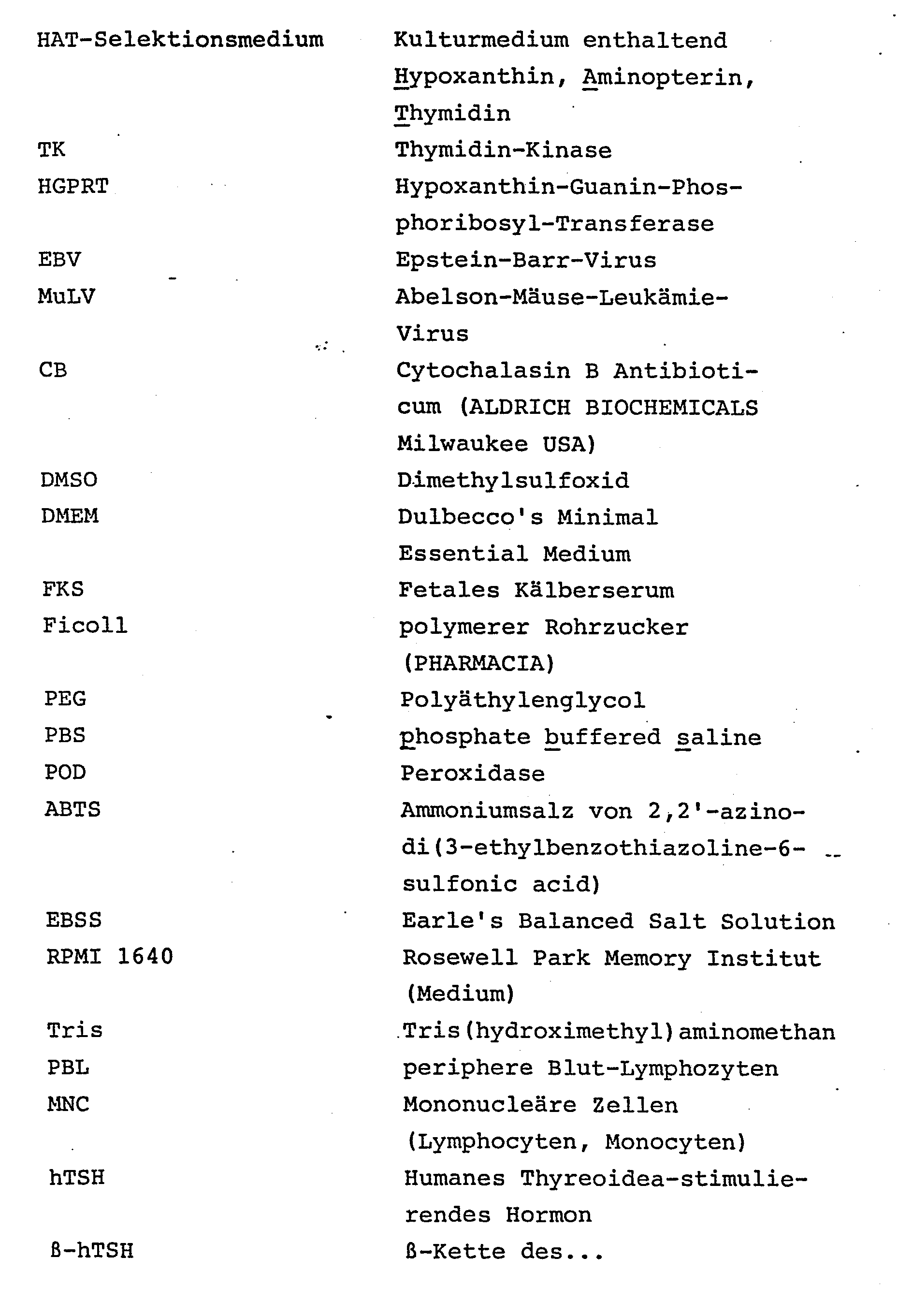
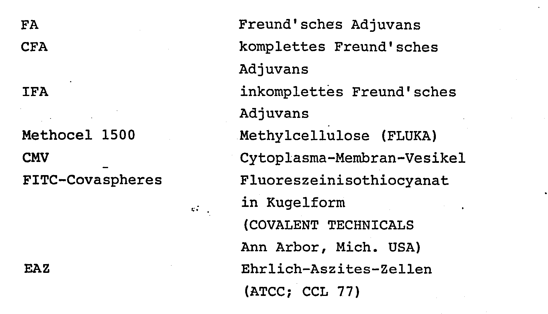
A Herstellung von Karyoplasten und Cytoplasten aus Maus-Myelomzellen der Linie P3X63 Ag 8.653 ATCC No-CRL-1580 (analog der in Wigler, M.H. and Weinstein, I.B.: A preparative method for obtaining enucleated mammalian cells. Biochem. Biophys. Res. Comm. 63, 669-674 (1975) -beschriebenen Methode).A Production of karyoplasts and cytoplasts from mouse myeloma cells of the line P3X63 Ag 8.653 ATCC No-CRL-1580 (analogous to that in Wigler, MH and Weinstein, IB: A preparative method for obtaining enucleated mammalian cells. Biochem. Biophys. Res. Comm. 63, 669-674 (1975) described method).
A.1 Material Cytochalasin B (CB, Aldrich Biochemicals, Milwaakee, USA) wurde in Dimethylsulfoxid (DMSO, Merck) gelöst (2 mg/ml) und als Stammlösung bei 4 °C gelagert.A.1 Material cytochalasin B (CB, Aldrich Biochemicals, Milwaakee, USA) was dissolved in dimethyl sulfoxide (DMSO, Merck) (2 mg / ml) and stored as a stock solution at 4 ° C.
Ficoll-400 (Pharmacia; polymerer Rohrzucker) wurde in redestilliertem Wasser gelöst (1 g/ml), autoklaviert und als 50 %ige Stammlösung bei -20 °C gelagert.Ficoll-400 (Pharmacia; polymeric cane sugar) was dissolved in redistilled water (1 g / ml), autoclaved and stored as a 50% stock solution at -20 ° C.
Einfach- und doppelkonzentriertes Dulbecco's Minimum Essential Medium (DMEM), Fetales Kälberserum (FKS), L-Glutamin (200 mmol/1), Streptomycin-Penicillin von Boehringer Mannheim. Cellulose-Nitratröhrchen wurden durch UV-Bestrahlung sterilisiert.Single and double concentrated Dulbecco's Minimum Essential Medium (DMEM), Fetal Calf Serum (FKS), L-Glutamine (200 mmol / 1), Streptomycin Penicillin from Boehringer Mannheim. Cellulose nitrate tubes were sterilized by UV radiation.
Myelomzell-Linie Ag 8.653 ATCC CRL-1580: Die Linie ist in Kearney, J.F. et al.: A new mouse myeloma cell line that hat lost immunoglobulin expression but permits the construction of antibodysecreting hybrid cell lines. J. Immunol. 123, 1548-1550 (1979) beschrieben. Sie ist Azaguinin- resistent, HAT-sensitiv und synthetisiert weder H- noch L-Ig-Ketten. Sie wird in DMEM + 15 % FKS + Glutamin + Penicillin-Streptomycin + Pyruvat (= DMEM-Vollmedium) bei 37 °C in 7 % C02-Atmosphäre gehalten.Myeloma cell line Ag 8.653 ATCC CRL-1580: The line is in Kearney, JF et al .: A new mouse myeloma cell line that has lost immunoglobulin expression but permits the construction of antibodysecreting hybrid cell lines. J. Immunol. 123, 1548-1550 (1979). It is azaguinine-resistant, HAT-sensitive and does not synthesize H or L-Ig chains. It is kept in DMEM + 15% FCS + glutamine + penicillin-streptomycin + pyruvate (= DMEM complete medium) at 37 ° C in a 7% C0 2 atmosphere.
A.2 Methoden Enukleierung: 8 x 10⁷ Ag 8.653-Zellen wurden 5 min bei 103 UpM zentrifugiert und in 12 ml einer 12,5 %igen Ficoll-DMEM-CB-DMSO-Lösung so lange resuspendiert, bis eine Zellklumpen-freie Suspension hergestellt war. Je 3 ml der Zellsuspension wurden auf 4, 12 Stunden vorher präparierte Ficoll-Gradienten, geschichtet und mit 2 ml Ficoll-freier DMEM-CB-DMSO-Lösung überschichtet. Die Röhrchen mit den Gradienten wurden in einer Ultrazentrifuge 60 Minuten lang bei 25.000 UpM (31 °C) zentrifugiert.A.2 Methods nucleation: 8 x 10 7 Ag 8,653 cells were centrifuged for 5 min at 10 3 rpm and resuspended in 12 ml of a 12.5% Ficoll-DMEM-CB-DMSO solution until a cell lump-free one Suspension was made. 3 ml portions of the cell suspension were layered on 4, 12 hours previously prepared Ficoll gradients and covered with 2 ml Ficoll-free DMEM-CB-DMSO solution. The gradient tubes were centrifuged in an ultracentrifuge for 60 minutes at 25,000 rpm (31 ° C).
Nach Beendigung der Zentrifugation wurden die makroskopischen sichtbaren Fraktionen ("Banden") mit Hilfe einer Injektionsspritze mit langer Kanüle von oben her getrennt gesammelt, in je 20 ml Kulturmedium (DMEM ohne Zusätze) verdünnt, durch Zentrifugation sedimentiert und in frischem DMEM resuspendiert.After centrifugation was complete, the macroscopic visible fractions ("bands") were collected separately from above using a syringe with a long cannula, each in 20 ml Culture medium (DMEM without additives) diluted, sedimented by centrifugation and resuspended in fresh DMEM.
Folgende 4 Fraktionen wurden erhalten:
- a) Zell-Debris (an der Grenze zwischen 0 und 12,5% Ficoll),
- b) kernlose Cytoplasten im Bereich von 15 bis 16 % Ficoll),
- c) Kerne ohne erkennbaren Plasmasaum und ca. 2 % kernlose "Zellen" an der Grenze zwischen 17 und 25 % Ficoll,
- d) kernhaltige, nach morphologischen Kriterien intakte Zellen mit gut erkennbarem Plasmasaum und wenige Kerne ohne Plasmasaum als Sediment auf dem Röhrchenboden.
- a) cell debris (on the border between 0 and 12.5% Ficoll),
- b) seedless cytoplasts in the range from 15 to 16% Ficoll),
- c) nuclei without a recognizable plasma margin and approx. 2% coreless "cells" at the border between 17 and 25% Ficoll,
- d) nucleated, intact cells according to morphological criteria with clearly recognizable plasma fringing and a few nuclei without plasma fringing as sediment on the tube bottom.
Die Zellzählung ergab, daß von den 8 x 10⁷ Ag 8.653-Zellen in b) 1,25 x 10⁶ Cytoplasten, in c) 4 x 10⁶ Karyoplasten und in d) 1,1 x 10. mutmaßlich intakte Zellen enthalten waren.The cell count showed that of the 8 x 10 7 A g 8,653 cells in b) 1.25 x 10 6 cytoplasts, in c) 4 x 10 6 karyoplasts and in d) 1.1 x 10 contained presumably intact cells .
B) Fusion von Maus-Milz-Zellen mit isolierten Myelom-Karyoplasten, -Cytoplasten und -Sedimentzellen aus Versuch A.B) Fusion of mouse spleen cells with isolated myeloma karyoplasts, cytoplasts and sediment cells from experiment A.
Fusionsmittel: 20 g Polyethylenglycol (PEG-4000) wurden im Autoklaven geschmolzen, auf 56 °C abgekühlt und bei dieser Temperatur mit 20 ml DMEM vermischt. HAT-Selektionsmedium: Zu DMEM-Vollmedium wurden Aminopterin (4 x 10⁻⁷ M), Thymidin (1 x 10⁻⁴ M) und Hypoxanthin (3,1 x 10⁻⁵ M) gegeben.Fusion agent: 20 g of polyethylene glycol (PEG-4000) were melted in an autoclave, cooled to 56 ° C. and mixed with 20 ml of DMEM at this temperature. H AT selection medium: Aminopterin (4 x 10- 7 M), thymidine (1 x 10- 4 M) and hypoxanthine (3.1 x 10 -5 M) were added to the DMEM complete medium.
Kulturgefäße: Tissue Culture Cluster 24 und Cluster 96 der Firma Costar, Cambridge, Mass., USA.Culture vessels: Tissue Culture Cluster 24 and Cluster 96 from Costar, Cambridge, Mass., USA.
Fusionen: Die im Versuch A präparierten Fraktionen b), c) und d) wurden in getrennten Ansätzen mit Milzzellen im Verhältnis 10:1 vermischt und durchFusions: The fractions b), c) and d) prepared in experiment A were mixed in separate batches with spleen cells in a ratio of 10: 1 and passed through
Zentrifugieren sedimentiert. Die überstehende Flüssigkeit wurde sorgfältig entfernt. Zu dem Sediment wurden 0,8 ml 50 %ige PEG-Lösung (bei 37 °C, gleichmäßig über 1 Minute verteilt, unter ständigem, sanften Schütteln) und dann 5 ml DMEM (bei Raumtemperatur, gleichmäßig über 5 Minuten) gegeben. Nach Zugabe von weiteren 20 ml DMEM wurden die Zellen sedimentiert, in frischem DMEM-Vollmedium (5 ml) resuspendiert, und auf je 10, mit "feedercells" beschichte 24er Costar-Tüpfel Gewebskulturgefäße verteilt. Die Einzelkulturen wurden am Tag 1, 2, 3, 5, 7, 10, 13 mit DMEM-Vollmedium "gefüttert".Centrifuged sedimented. The supernatant was carefully removed. 0.8 ml of 50% PEG solution (at 37 ° C., evenly distributed over 1 minute, with constant, gentle shaking) and then 5 ml of DMEM (at room temperature, evenly over 5 minutes) were added to the sediment. After the addition of a further 20 ml of DMEM, the cells were sedimented, resuspended in fresh DMEM complete medium (5 ml), and tissue tissue vessels were distributed into 10 24-well Costar spots coated with “feedercells”. The individual cultures were "fed" on day 1, 2, 3, 5, 7, 10, 13 with full DMEM medium.
Feeder-cells ((Bauchhöhlen-Makrophagen): Am Tag vor der Fusion wurden Inzuchtmäuse (Balb/c) durch Streckung getötet. Unter sterilen Bedingungen wurden 4 bis 5 ml PBS in die Bauchhöhle gespritzt und nach 1 Minute wieder abgesaugt. Die ausgespülten Zellen wurden in DMEM gewaschen, in Vollmedium auf eine Dichte von 2 x 10⁵ Zellen pro ml suspendiert und in 0,5 ml-Portionen auf 24er Costar-Tüpfel verteilt.Feeder-cells (invertebral macrophages): Inbred mice (Balb / c) were sacrificed by extension the day before the fusion and 4 to 5 ml of PBS were injected into the abdominal cavity under sterile conditions and aspirated again after 1 minute. The rinsed-out cells were washed in DMEM, suspended in full medium to a density of 2 × 10 5 cells per ml and distributed in 0.5 ml portions on 24 pieces of Costar.
Milzzellen: Einer Balb/c-Maus wurde unmittelbar vor Fusion unter aseptischen Bedingungen die Milz exstirpiert und deren Zellen in DMEM suspendiert. Zellaggregate und Gewebsstücke wurden mit Mullgaze abfiltriert.Spleen cells: A Balb / c mouse was immediately excised from the spleen under aseptic conditions and its cells suspended in DMEM. Cell aggregates and pieces of tissue were filtered off with gauze gauze.
Elisa auf Maus-Immunoglobulin: Mikrotiterplatten wurden mit Maus-Ig-Antikörpern vom Schaf (IgG-Fraktion; 10 µg/ml 0,9 %ige NaCl- Lösung; 150 µl Antikörper-Lösung pro Tüpfel) beschichtet. Je 100 µl Kulturüberstand wurden in die beschichteten Tüpfel pipettiert und eine Stunde bei Raumtemperatur inkubiert. Nach Absaugen der Überstände und zweimaligem Waschen wurden die Tüpfel mit 100 µl anti-Maus-Ig-POD-Konjugat-Lösung (gleicher Antikörper wie oben; kovalent verbunden mit Meerrettich-Peroxidase) beschickt und eine Stunde bei Raumtemperaturinkubiert. Nach dreimaligem Waschen wurden pro Tüpfel 100 µl Substrat- Lösung (ABTS) pipettiert und die Farbentwicklung photometrisch bestimmt.Elisa on mouse immunoglobulin: microtiter plates were coated with mouse Ig antibodies from sheep (IgG fraction; 10 µg / ml 0.9% NaCl solution; 150 µl antibody solution per well). 100 µl of culture supernatant were pipetted into the coated spot and incubated for one hour at room temperature. After the supernatants had been suctioned off and washed twice, the spots were charged with 100 μl of anti-mouse-Ig-POD conjugate solution (same antibody as above; covalently linked to horseradish peroxidase) and incubated for one hour at room temperature. After washing three times, 100 μl of substrate solution (ABTS) were pipetted into each well and the color development was determined photometrically.
Die gemäß A präparierten Cytoplast-, Karyoplast-und Sediment-Fraktionen wurden parallel mit Milz- zellen einer Balb/c-Maus fusioniert und auf je 10 1-ml-Kulturen verteilt. Je 5 der Teilkulturen wurden ohne (Platte I) und je 5 mit HAT-Zusatz (Platte II) gefüttert.The cytoplast, karyoplast and sediment fractions prepared according to A were fused in parallel with spleen cells of a Balb / c mouse and distributed over 10 1 ml cultures in each case. 5 of each subculture were fed without (plate I) and 5 each with HAT addition (plate II).
Bis zum Tag 21 nach Fusion (n.F.) war in keinem der Tüpfel Wachstum von lymphoiden Zellen makroskopisch oder mikroskopisch nachweisbar, mit Ausnahme der Tüpfel 4A, 4B, 4C, 3C und 3D auf Platte I, die das Fusionat der Sedimentfraktion ohne HAT-Medium enthielten. In diesen Tüpfeln waren bereits 5 Tage nach Fusion (n.F.) Kolonien erkennbar, die sich rapide vergrößerten. Am Tag 8 n.F. wurde in diese Tüpfel HAT-Medium gegeben: innerhalb von 4 Tagen waren daraufhin alle sichtbaren Kolonien abgestorben.Up to day 21 after fusion (nF), growth of lymphoid cells was not detectable macroscopically or microscopically in any of the wells, with the exception of wells 4A, 4B, 4C, 3C and 3D on plate I, which contained the fusion of the sediment fraction without HAT medium . In these spots, colonies were already visible 5 days after fusion (n.F.), which grew rapidly. On day 8 a.m. HAT medium was added to this dot: all visible colonies had died within 4 days.
Ab Tag 27 n.F. wurden zunächst vereinzelt, dann in nahezu allen Tüpfeln Kolonien sichtbar, die aus großen, kugeligen, transparenten, nicht-adhärentwachsenden Zellen bestanden. Am Tag 65 n.F. waren mit Ausnahme von I-3B, II-lA, II-4A alle Tüpfel mit multiplen Kolonien von lymphoiden Zellen besetzt. Die Prüfung der Kulturüberstände auf Gehalt an Maus-Ig an diesem Tag ergab, wie die Zusammen-__ stellung in Tabelle 1 zeigte, positive bis stark positive Werte im ELISA in allen Teilkulturen mit Ausnahme der obengenannten Kolonie-freien Tüpfel:


- a) Die Fusion von Karyoplasten mit Maus-Milz-Zellen führte zu in vitro vermehrbaren, Immunglobulin-sezernierenden Zellen. Obwohl nur ein kleiner Teil des Cytoplasmas des malignen Partners in das Hybrid eingebracht wurde, kam es nicht zu der möglichen Extinktion des Merkmals "permanentes Wachstum".
- b) Die mit Karyoplasten erzeugten Hybridzellen synthetisierten und sezernierten Maus-Immunglobulin in "Hybridoma"-Quantitäten. Das Fehlen des Cytoplasma-Anteils der Ag 8.653 beeinträchtigte demnach die Produktions- und Sekretionsfähigkeit der Karyoplast-Milzzell-Hybride nicht.
- c) Aus der Fusion von Cytoplasten mit Milzzellen gingen ebenfalls Zellklone hervor, die in vitro proliferierten und Antikörper sezernierten. Eine befriedigende Erklärung ist für dieses besonders überraschende Phänomen z. Z. nicht zu geben.
- a) The fusion of karyoplasts with mouse spleen cells led to in vitro reproducible, immunoglobulin-secreting cells. Although only a small part of the malignant partner's cytoplasm was introduced into the hybrid, there was no possible extinction of the characteristic "permanent growth".
- b) The hybrid cells produced with karyoplasts and synthesized and secreted mouse immunoglobulin in "hybridoma" quantities. The lack of the cytoplasmic part of Ag 8.653 did not impair the production and secretion ability of the karyoplast-spleen cell hybrids.
- c) The fusion of cytoplasts with spleen cells also gave rise to cell clones that proliferated in vitro and secreted antibodies. A satisfactory explanation for this particularly surprising phenomenon is e.g. Currently not to be given.
Zellfragmentierung mittels Glycerin-Lyse und Fusion mit humanen Blut-LymphocytenCell fragmentation using glycerol lysis and fusion with human blood lymphocytes
Earle's Balanced Salt Solution (EBSS), Kulturmedium APMI 1640, Fetales Kälberserum (FKS) von Boehringer Mannheim. 8-Azaguanin (8-Ag) von Serva (Heidelberg), Agar (Bacto-Agar 1614) von Difco (Fa. Hedinger KG, Stuttgart). Die humane Plasmacytom-Linie HS SULTAN ATCC CRL-1484 wurde als kryopräserviertes Zellmaterial nach ATCC-Vorschrift aufgetaut und in Kultur genommen.Earle's Balanced Salt Solution (EBSS), culture medium APMI 1640, fetal calf serum (FKS) from Boehringer Mannheim. 8-Azaguanine (8-Ag) from Serva (Heidelberg), Agar (Bacto-Agar 1614) from Difco (from Hedinger KG, Stuttgart). The human plasmacytoma line HS SULTAN ATCC CRL-1484 was thawed as a cryopreserved cell material according to the ATCC specification and taken in culture.
Nach dem in Proc. Natl. Acad. Sci. USA. 71, 2679-2683 (1974) beschriebenen Verfahren wurden 8 x 10⁷ HS Sultan-Zellen in 100 ml RPMI 1640-Vollmedium, das 20 µM 8-Ag enthielt, 48 Stunden lang kultiviert. Die überlebenden Zellen wurden in 10 ml RPMI 1640-Vollmedium ohne 8-Ag aufgenommen und über 10 Tage vermehrt.According to the in Proc. Natl. Acad. Sci. UNITED STATES. 71, 2679-2683 (1974), 8 × 10 7 HS Sultan cells were cultured in 100 ml RPMI 1640 complete medium containing 20 μM 8-Ag for 48 hours. The surviving cells were taken up in 10 ml RPMI 1640 complete medium without 8 Ag and multiplied over 10 days.
Die Zellen wurden sodann auf Soft-Agar-Platten (COFFINO, P. et al: Proc. Natl. Acad. Sci. USA. 68, 219-223 (1971)), die mit RPMI 1640-Vollmedium + 20 µM 8-Ag hergestellt worden waren, ausgesät (ca. 500 Zellen pro Petrischale) und im C02-Inkubator bebrütet. Nach 9 Tagen wurden isoliert wachsende Kolonien steril von der Agaroberfläche entnommen und in RPMI 1640-Vollmedium + 8-Ag vermehrt. Ein Klon (als HS-SULTAN-8Ag-R1 bezeichnet), der mit einer Verdopplungszeit von ca. 20 Stunden wuchs, wurde für den folgenden Versuch verwendet. Die HS-Rl-Zellen waren HAT-sensitiv: Zellen, die in einer Dichte von 1 bis 5 x 10⁵ pro ml HAT-Medium (RPMI 1640-Vollmedium mit 0,1 mM Hypoxanthin, 400 nM Aminopterin, 31 µM Thymidin) kultiviert wurden, vermehrten sich nicht und starben innerhalb von 7 Tagen vollständig ab.The cells were then placed on soft agar plates (COFFINO, P. et al: Proc. Natl. Acad. Sci. USA. 68, 219-223 (1971)), which were filled with RPMI 1640 complete medium + 20 μM 8-Ag had been produced, sown (approx. 500 cells per Petri dish) and incubated in the C0 2 incubator. After 9 days, isolated growing colonies were removed sterile from the agar surface and grown in RPMI 1640 complete medium + 8-Ag. A clone (designated HS-SULTAN-8Ag-R1) that grew with a doubling time of approximately 20 hours was used for the following experiment. The HS-R1 cells were HAT-sensitive: cells which were cultured in a density of 1 to 5 × 10 5 per ml of HAT medium (RPMI 1640 complete medium with 0.1 mM hypoxanthine, 400 nM aminopterin, 31 μM thymidine) did not reproduce and died completely within 7 days.
Humane Lymphocyten (aus dem peripheren Blut: PBL): 300 ml Venenblut wurden steril in Heparin-Lösung (2 U/ml Blut) gesammelt und die Fraktion der mononukleären Zellen (MNC: Lymphocyten, Monocyten) mit Standard-Methoden isoliert. 3 x 10⁸ MNC wurden in 100 ml RPMI 1640 x 10 % FKS suspendiert und zur Abtrennung der Monocyten 24 h in Kulturgefäßen bei 37 °C in 5 % C02-Atmosphäre inkubiert.Human lymphocytes (from the peripheral blood: PBL): 300 ml of venous blood were sterile collected in heparin solution (2 U / ml blood) and the fraction of the mononuclear cells (MNC: lymphocytes, monocytes) was isolated using standard methods. 3 x 10 8 MNC were suspended in 100 ml RPMI 1640 x 10% FCS and incubated for 24 h in culture vessels at 37 ° C in a 5% CO 2 atmosphere to separate the monocytes.
Fragmentierung von HS-R1: Die Zellen wurden nach (Jett, M. et al.: Isolation and characterization of plasma membranes and intact nuclei from lymphoid cells. J. Biol. Chem. 252, 2134-2142 (1977)) mit Glycerin beladen und durch Inkubation in 10 mM Tris-HCl-Puffer lysiert. Die Kerne wurden durch Zentrifugation mit 200 g (10 Minuten, 4 °C) von den Membranvesikeln abgetrennt, die ihrerseits durch Zentrifugation bei 5000 g (40 Minuten, 4 °C) sedimentiert wurden.Fragmentation of HS-R1: The cells were loaded with glycerol according to (Jett, M. et al .: Isolation and characterization of plasma membranes and intact nuclei from lymphoid cells. J. Biol. Chem. 252, 2134-2142 (1977)) and lysed by incubation in 10 mM Tris-HCl buffer. The cores were separated from the membrane vesicles by centrifugation at 200 g (10 minutes, 4 ° C.), which in turn were sedimented by centrifugation at 5000 g (40 minutes, 4 ° C.).
Fusion: Ca. 1 x 10⁸ 729 HS-R1-Kerne wurden mit Human-Lymphocyten in RPMI 1640 im Verhältnis 1:1 suspendiert und, wie in Beispiel 1, B.2 beschrieben, mittels PEG fusioniert.Fusion: Approx. 1 x 10 8 729 HS-R1 nuclei were suspended with human lymphocytes in RPMI 1640 in a ratio of 1: 1 and, as described in Example 1, B.2, fused using PEG.
Das Sediment von Membranvesikeln aus etwa 1 x 10⁸ HS-R1-Zellen wurde mit einer Suspension von 1 x 10⁷ Human- Lymphocyten überschichtet und mittels PEG fusioniert.The sediment of membrane vesicles from approximately 1 x 10 8 HS-R1 cells was overlaid with a suspension of 1 x 10 7 human lymphocytes and fused using PEG.
In einem Kontrollansatz wurden ca. 2 x 10⁷ HS-Rl-Kerne in RPMI 1640-Vollmedium (ohne HAT) aufgenommen und in 4 24er Costar-Tüpfeln im C02-Inkubator in Kultur genommen.In a control batch, approx. 2 x 10 7 HS-Rl cores were taken up in RPMI 1640 full medium (without HAT) and in culture in 4 24-wells of Costar in the C0 2 incubator.
Alle Fusionate und die Kontroll-Kultur wurden auf Maus-Bauchhöhlen-Makrophagen wie in Beispiel 1 beschrieben, als feeder- cells kultiviert.All fusionates and the control culture were cultivated on mouse abdominal macrophages as described in Example 1 as feeder cells.
Nachweis von Human-Ig in Kulturüberständen: Mikrotiter-ELISA wie in Beispiel 1, B.2 beschrieben. Zur Beschichtung wurde immunadsorptiv gereinigtes anti-human-Ig vom Schaf verwandt. Die gleiche AK-Präparation wurde zur Herstellung des anti-human-Ig-POD-Konjugats verwandt. Der Schaf-AK reagierte mit allen menschlichen Ig-Klassen und zeigte keine Kreuzreaktivität mit bovinem oder murinem Ig.Detection of human Ig in culture supernatants: microtiter ELISA as described in Example 1, B.2. Immunosorptively purified sheep anti-human Ig was used for the coating. The same AK preparation was used to make the anti-human-Ig-POD conjugate. The sheep AK reacted with all human Ig classes and showed no cross-reactivity with bovine or murine Ig.
Fragmentierung durch Glycerin-Tris-HCl-Lyse: Die Behandlung zerlegte HS-R1-Zellen in eine kernhaltige Fraktion, die bei 200 g sedimentierte und in eine kernlose Cytoplasma-Membran-Vesikel-Fraktion, die bei 5000 g sedimentierbar war. Unter dem Mikroskop waren in keiner der beiden Fraktionen intakte HS-R1-Zellen erkennbar. Die Kerne waren mit mehr oder weniger, unregelmäßig begrenzten Cytoplasma-Fetzen umgeben. Die. Auszählung ergab eine Kern-Ausbeute von 85 %. Die Vesikel-Fraktion enthielt neben wenigem Debris massenhaft 0,5 bis 2 µm große Vesikel ohne erkennbare Kernbestandteile.Fragmentation by glycerol-Tris-HCl lysis: The treatment split HS-R1 cells into a nucleated fraction, which sedimented at 200 g and into a coreless cytoplasmic membrane vesicle fraction, which was sedimentable at 5000 g. Under the microscope, intact HS-R1 cells were not visible in either of the two fractions. The nuclei were surrounded by more or less, irregularly delimited cytoplasm scraps. The. Counting showed a core yield of 85%. In addition to a small amount of debris, the vesicle fraction contained a mass of 0.5 to 2 µm large vesicles without recognizable core components.
Die Kultur von 2 x 10 Kernen in RPMI-Vollmedium (ohne HAT) führte in einem Beobachtungszeitraum von 12 Wochen zu keinem Wachstum von HS-R1-Zellen.The culture of 2 × 10 nuclei in RPMI complete medium (without HAT) did not lead to growth of HS-R1 cells in an observation period of 12 weeks.
Kultur der Fusionate: Die Kern-Lymphocyten- und Cytoplasma-Vesikel-Lymphocyten-Fusionate wurden auf 96 bzw. 10 24er Costar-Tüpfel verteilt und je zur Hälfte mit und ohne HAT-Zusatz in RPMI-Vollmedium auf Maus-Makrophagen kultiviert. Ab der zweiten Woche nach Fusion wurden Kolonien von lymphoiden Zellen sichtbar, die kontinuierlich an Größe zunahmen.Culture of the fusionates: The core-lymphocyte and cytoplasm-vesicle-lymphocyte fusions were distributed to 96 and 10 24-wells of Costar, respectively, and half cultivated on mouse macrophages in RPMI complete medium with and without HAT addition. From the second week after fusion, colonies of lymphoid cells became visible and continued to grow in size.
Produktion von humanem Immunglobulin: Am Tag 21 nach Fusion wurden Kulturüberstände (am Tag 19 war ein kompletter Medium-Wechsel vorgenommen worden) auf Gehalt an menschlichem Immunglobulin geprüft. Die Ergebnisse zeigen die Tabellen 2a und 2b.
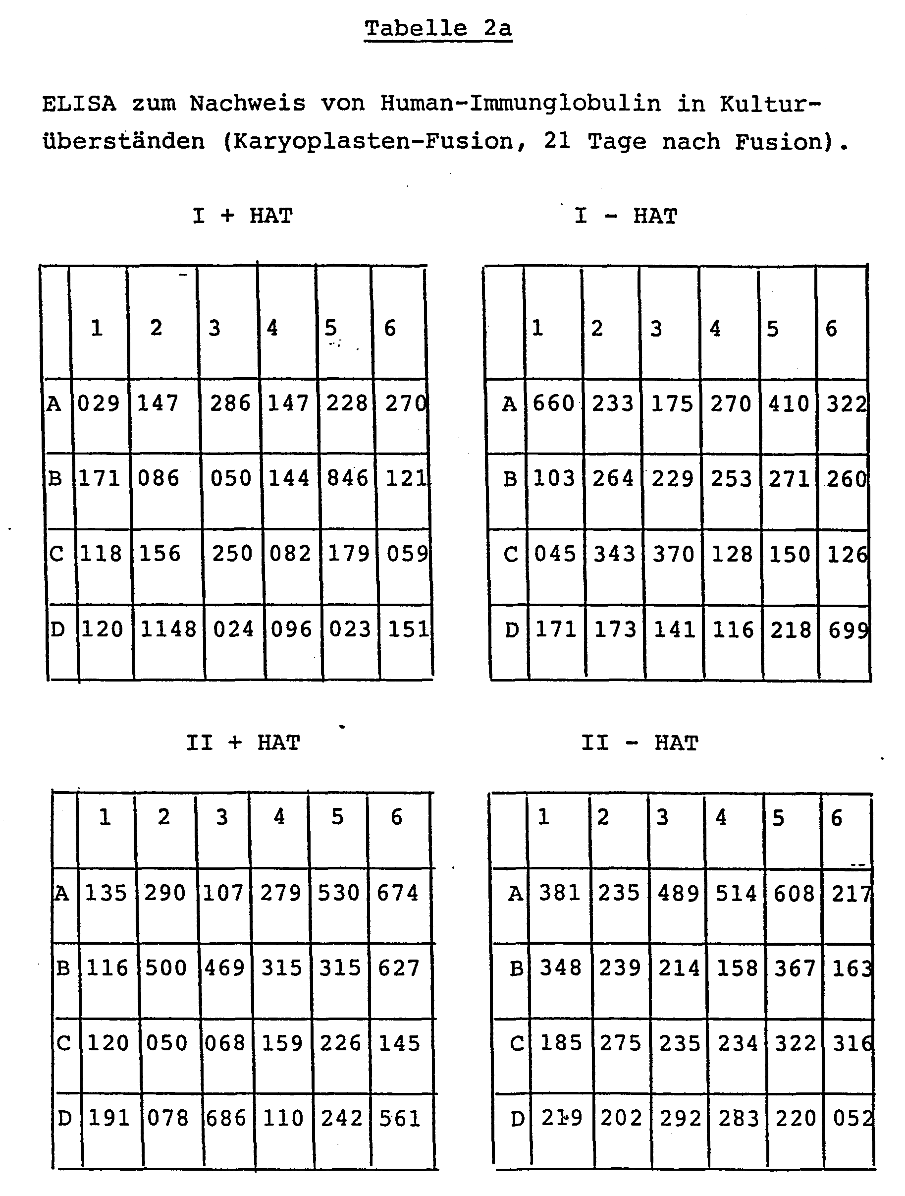

Setzt man die Extinktionswerte von 0 bis 99 als negativ bis zweifelhaft, die Werte von 100 bis 200 als positiv, die Werte >200 als stark positiv an, dann ergibt sich für die durch Kernfusion erzeugten Kulturen folgende Verteilung:If one takes the extinction values from 0 to 99 as negative to doubtful, the values from 100 to 200 as positive, the values> 200 as strongly positive, then the following distribution results for the cultures produced by nuclear fusion:

Fusionate,.die mit Lyse-Fraktionen hergestellt werden, können ohne HAT-Selektion kultiviert werden. Das Ver- fahren ist sehr viel weniger arbeitsaufwendig als die Cytochalasin-B-Extrusion und liefert fusionsfähiges Material in hoher Ausbeute. Eine klare Auftrennung in "Kern-" und "Cytoplasma-Membran"-Fraktionen wird mit keinem der beiden Verfahren erreicht. Sowohl die überwiegend Kern-Material enthaltende wie die überwiegend Cytoplasma-Membran-Vesikel enthaltende Fraktion erzeugt nach Fusion mit menschlichen Lymphocyten aus dem Blut in vitro vermehrungsfähige, AK-produzierende Zell-Klone.Fusionates made with lysis fractions can be cultured without HAT selection. The process is much less labor-intensive than cytochalasin B extrusion and delivers fusionable material in high yield. A clear separation into "core" and "cytoplasmic membrane" fractions is not achieved with either method. After the fusion with human lymphocytes, both the fraction containing predominantly core material and the fraction containing predominantly cytoplasmic membrane vesicles generates reproductive, AK-producing cell clones from the blood.
Der Parallel-Ansatz der Kern-Lymphocyten-Hybride mit und ohne HAT-Zusatz dokumentiert die negativen Auswirkungen des Selektionsmediums: Mit HAT sind 23 % der Primärkulturen Ig-negativ und nur 40 % stark positiv; ohne HAT sind über 70 % stark positiv und nur 4 % Ig-negativ.The parallel approach of nuclear-lymphocyte hybrids with and without HAT supplement documents the negative impact of the selection medium: With HAS 23% of P rimärkulturen Ig negative and only 40% strongly positive; Without HAT, over 70% are strongly positive and only 4% are Ig negative.
Fusion von Milzzellen einer immunisierten Maus mit nativen und fragmentierten.Myelomzellen Ag 8.653Fusion of spleen cells of an immunized mouse with native and fragmented. Myeloma cells Ag 8.653
Humanes Thyreoidea-stimulierendes Hormon (hTSH) und dessen isolierte ß-Kette (ß-hTSH) stammten von Boehringer Mannheim. Komplettes und Inkomplettes Freund'sches Adjuvans (CFA, IFA) von Difco, Methocel 1500 von Fluka und FITC-Covaspheres von Covalent Tech. Co., Ann. Arbor, Michigan, USA.Human thyroid stimulating hormone (hTSH) and its isolated ß chain (ß-hTSH) were from Boehringer Mannheim. Complete and Incomplete Freund's Adjuvant (CFA, IFA) from Difco, Methocel 1500 from Fluka and FITC-Covaspheres from Covalent Tech. Co., Ann. Arbor, Michigan, United States.
Immunisierung: Balb/c-Mäuse wurden mit ßhTSH (40 µg in CFA, intraperitoneal) primär immunisiert (Tag 1), und am Tag 196 mit hTSH (50 µg in IFA, i.p.), am Tag 266 mit hTSH ohne Adjuvans i.p. sowie am Tag 294 mit hTSH intravenös "geboostert".Immunization: Balb / c mice were primarily immunized with ßhTSH (40 µg in CFA, intraperitoneally) (day 1), and on day 196 with hTSH (50 µg in IFA, i.p.), on day 266 with hTSH without adjuvant i.p. and intravenously boosted with hTSH on day 294.
Milzzellen (ca. 1 x 10⁸) wurden von einer der immunisierten Mäuse 3 Tage nach der letzten Booster-Immunisierung, wie in Beispiel 1, B.2 beschrieben, gewonnen und je zur Hälfte für zwei Fusionen verwandt.Spleen cells (approx. 1 x 10 8 ) were obtained from one of the immunized mice 3 days after the last booster immunization, as described in Example 1, B.2, and half of them were used for two fusions.
Fusion 1: 5 x 10⁷ Milzzellen und 1 x 10⁷ Ag 8.653-Zellen wurden vermischt, wie in Beispiel 1, B.2 beschrieben, fusioniert und in 48 24er Tüpfel mit Maus-Makrophagen-Zellen in HAT-haltigem DMEM-Vollmedium kultiviert.Fusion 1: 5 × 10 7 spleen cells and 1 × 10 7 Ag 8,653 cells were mixed, as described in Example 1, B.2, fused and cultivated in 48 spots of 24 with mouse macrophage cells in HAT-containing DMEM full medium .
Fusion 2: 1 x 10⁷ Ag 8.653-Zellen wurden wie in Beispiel 2 beschrieben, durch schrittweise Behandlung mit Glycerin und 10 mM Tris-HCl-Puffer lysiert. Die Cytoplasma-Membran-Vesikel-(CMV-)Fraktion wurde pelletiert (5.000 g, 40 Minuten, 4 °C). Die Kern-Fraktion wurde mit 7 x 10⁷ Milzzellen vermischt, durch Zentrifugation auf das CMV-Sediment geschichtet und mittels PEG nach dem Standardverfahren, wie in Beispiel 1, B.2 beschrieben, fusioniert. Das Fusionat wurde im DMEM-Vollmedium auf 24 24er Costar-Tüpfeln mit Makrophagen erteilt und ohne HAT-Zusätze kultiviert.Fusion 2: 1 × 10 7 Ag 8,653 cells were lysed as described in Example 2 by stepwise treatment with glycerol and 10 mM Tris-HCl buffer. The cytoplasmic membrane vesicle (CMV) fraction was pelleted (5,000 g, 40 minutes, 4 ° C). The core fraction was mixed with 7 × 10 7 spleen cells, layered by centrifugation on the CMV sediment and fused by means of PEG according to the standard method as described in Example 1, B.2. The fusion was given in full DMEM medium on 24 Costar 24 spots with macrophages and cultured without HAT additives.
Antigenspezifische Markierungen von Hybridzellen und Klonierung: (FITC-) Covaspheres wurden nach der allgemeinen Vorschrift des Herstellers mit hTSH kovalent beschichtet (TSH-CS) und in 5 ‰ Natriumazid-Lösung gelagert. Nach dem in PARKS, D.R. et al: Proc. Natl. Acad. Sci. USA 76, 1982-1966 (1979) beschriebenen Verfahren wurden die Zellen mit den beschichteten Covaspheres markiert und mit Hilfe eines Cytofluorographen große, Fluoreszenz-positive Zellen einzeln in Tüpfel und 96er Costar-Platten "abgelegt". Die Tüpfel waren 24 Stunden zuvor mit Maus-Makrophagen in DMEM-Vollmedium beschicht worden.Antigen-specific labeling of hybrid cells and cloning: (FITC-) covaspheres were covalently coated with hTSH (TSH-CS) according to the manufacturer's general instructions and stored in 5% sodium azide solution. After the in PARKS, D.R. et al: Proc. Natl. Acad. Sci. USA 76, 1982-1966 (1979), the cells were labeled with the coated covaspheres and, with the aid of a cytofluorograph, large, fluorescence-positive cells were individually “deposited” in wells and 96-well Costar plates. The spots had been coated with mouse macrophages in DMEM complete medium 24 hours previously.
ELISA für TSH-spezifische Antikörper: Beschichtung, Inkubation der Kulturüberstände, Substrat-Reaktion und Ablesung wie in Beispiel 1, B.2, beschrieben. Anstelle von anti-Maus-Ig-POD wurde TSH-POD-Konjugat verwendet. Als positive Kontrolle wurde Serum einer hTSH-hyperimmunisierten Maus, 10-3 verdünnt, eingesetzt; als Negativ-Kontrolle diente ein Klon, welcher Antikörper gegen ein nichtverwandtes Antigen bildete (Maus-anti-Digoxin).ELISA for TSH-specific antibodies: coating, incubation of the culture supernatants, substrate reaction and reading as described in Example 1, B.2. Instead of anti-mouse Ig-POD, TSH-POD conjugate was used. Serum from an hTSH hyperimmunized mouse, diluted 10 -3 , was used as a positive control; a clone, which produced antibodies against an unrelated antigen (mouse anti-digoxin), served as a negative control.
Sowohl in den Kulturen aus Fusion 1 (mit intakten Ag 8.653-Zellen) als auch denen aus Fusion 2 (mit Ag 8.653-Lyse-Fragmenten) waren am Tag 14 in allen Tüpfeln Kolonien von großen, lymphoiden Zellen erkennbar. Der mit Kulturüberständen vom Tag 14 durchgeführte ELISA zum Nachweis von TSH-spezifischen Antikörpern ergab die in Tabelle 3 zusammengefaßten Werte: In allen Teilkulturen wurde anti-TSH nachgewiesen.

Am Tag 15 wurden die nicht-adhärenten Zellen aus den Einzel-Tüpfeln von Fusion 1 und 2 herausgespült und in getrennten Ansätzen TSH-Antigen-spezifisch markiert (Grundlage: Hybridoma tragen in der Regel wie B-Lymphocyten einen Teil der von ihnen synthetisierten Antikörpermoleküle in der Zellmembran verankert, mit "nach außen" gerichteten Antigen-Bindungsstellen) und mit Hilfe des Zellsorters kloniert.On day 15, the non-adherent cells were rinsed out of the individual wells of fusion 1 and 2 and labeled in a separate batch specifically for TSH antigens (basis: hybridomas, like B lymphocytes, usually carry part of the antibody molecules they synthesized into them anchored to the cell membrane, with "outward" antigen binding sites) and cloned with the help of the cell sorter.
Fusion von Humanen PBL mit intakten und mit fragmentierten Ag 8.653-Zellen.Fusion of human PBL with intact and with fragmented Ag 8.653 cells.
Inaktiviertes Hepatitis-B-Surface-Antigen (HBsi; Biotest) wurde durch Immunadsorption von Serumproteinen gereinigt. ELISA zum Nachweis von humanen HBs-Antikörpern: Mikrotiter-Platten wurden mit gereinigtem HBsi (20 ug/ml 0,9%ige NaCl-Lösung) beschichtet. Inkubation der Kulturüberstände, Konjugat-und Substrat-Reaktion sowie Ablesung wie im Beispiel 1.B.2 beschrieben. Anstelle von Anti-Maus-Ig-POD wurde (Schaf-)Anti-Human-Ig-POD verwendet.Inactivated hepatitis B surface antigen (HB si ; Biotest) was purified from serum proteins by immunoadsorption. ELISA for the detection of human HB s antibodies: Microtiter plates were coated with purified HB si (20 µg / ml 0.9% NaCl solution). Incubate the culture supernatants, conjugate and substrate reaction and read as described in Example 1.B.2. Instead of anti-mouse Ig-POD, (sheep) anti-human Ig-POD was used.
HPBL von einem Spender mit hohem anti-HBs-Titer wurden aus 200 ml Venenblut durch Ficoll-Gradienten-Zentrifugation isoliert. Zur Anreicherung der B-Lymphocyten wurden die T-Zellen mit Schaferythrocyten (nach Standardverfahren) rosettiert und mittels einer zweiten Ficoll-Gradienten-Zentrifugation abgetrennt. Die Fraktion der nicht-rosettierenden Zellen (HPBL(B)) wurden in einer Dichte von 5 x 10⁷ Zellen in RPMI 1640 + 10 % autologem Plasma (30 min/56 °C-hitzeinaktiviert) mit ca. 10 µg HBsi im CO2-Inkubator kultiviert (Mediumwechsel alle 12 Stunden).HPBL from a high anti-HBs titer donor was isolated from 200 ml of venous blood by Ficoll gradient centrifugation. To enrich the B lymphocytes, the T cells were rosetted with sheep erythrocytes (according to standard procedures) and removed using a second Ficoll- Gradient centrifugation separated. The fraction of the non-rosetting cells (HPBL (B)) were in a density of 5 × 10 7 cells in RPMI 1640 + 10% autologous plasma (30 min / 56 ° C. heat-inactivated) with approx. 10 μg HBsi in CO 2 - Cultivated incubator (medium change every 12 hours).
Fusion 1: 1 x 10 der vorbehandelten HPBL(B) wurden mit 1 x 10⁷ Ag 8.653 vermischt und, wie in Beispiel 1, B.2 beschrieben, mittels PEG fusioniert. Das Fusionat wurde in RPMI 1640 + 10 % Humanplasma + HAT in 4 24er Costar-Tüpfeln kultiviert.Fusion 1: 1 x 10 of the pretreated HPBL (B) were mixed with 1 x 10 7 Ag 8.653 and, as described in Example 1, B.2, fused using PEG. The fusionate was cultivated in RPMI 1640 + 10% human plasma + HAT in 4 Costar 24 spots.
Fusion 2: 1,1 x 10⁸ Ag 8.653-Zellen wurden mittels Glycerin-Lyse fragmentiert. 1 x 10⁷ Ag 8.653-Kerne wurden mit 1 x 10⁷ HPBL(B) vermischt und auf das Sediment von Membran-Cytoplasma-Vesikeln (aus 1 x 10⁸ Ag 8.653-Zellen) geschichtet. Nach Fusion mit PEG wurde das Fusionat in 4 24er Costar-Tüpfeln in RPMI 1640 + 10 % Humanplasma (ohne HAT-Zusatz) kultiviert.Fusion 2: 1.1 x 10 8 Ag 8,653 cells were fragmented by glycerol lysis. 1 x 10 7 Ag 8.653 nuclei were mixed with 1 x 10 7 HPBL (B) and layered on the sediment of membrane cytoplasmic vesicles (from 1 x 10 8 A g 8,653 cells). After fusion with PEG, the fusion was cultivated in 4 24 wells of Costar in RPMI 1640 + 10% human plasma (without addition of HAT).
In allen Tüpfeln von Fusion 1 und 2 trat ab Tag 3 starkes Wachstum von sehr großen, adhärenten Zellen auf. Am Tag 5 wurde die Hauptmasse der nicht-adhärenten Zellen vorsichtig suspendiert und auf zwei neue 24er Costar-Tüpfel verteilt (z. B. A1 B1 und Cl). Am Tag 28 in Fusion 1: kleine Kolonien in A2 und A3, in den restlichen Tüpfeln nur adhärente Zellen; in Fusion 2: massives Wachstum multipler Kolonien in allen Tüpfeln mit Ausnahme von C3. Der mit Kulturüberständen vom Tag 35 durchgeführte ELISA zum Nachweis von humanen Immunglobulinen mit Spezifität gegen Hepatitis-B-Antigen ergab das in Tabelle 4 zusammengefaßte Ergebnis: die Fusion 1 (intakte Ag 8.653-Zellen, Kultur in HAT-Medium) lieferte keine positive Kultur; dagegen waren 5 von 12 Kulturen der Fusion 2 (Ag 8.653-Fragmente, Kultur in Normal-Medium) eindeutig Anti-HBs-positiv.From day 3 onwards, all of the spots in Fusion 1 and 2 showed strong growth of very large, adherent cells. On day 5, the main mass of the non-adherent cells was carefully suspended and divided into two new 24-wells of Costar (e.g. A1 B1 and Cl). On day 28 in Fusion 1: small colonies in A2 and A3, in the remaining Tü p feln only adherent cells; in Fusion 2: massive growth of multiple colonies in all wells except C3. The ELISA performed with culture supernatants from day 35 for the detection of human immunoglobulins with specificity against hepatitis B antigen revealed the result summarized in Table 4: Fusion 1 (intact Ag 8,653 cells, culture in HAT medium) gave no positive culture; in contrast, 5 of 12 cultures of fusion 2 (Ag 8,653 fragments, culture in normal medium) were clearly anti-HB s positive.
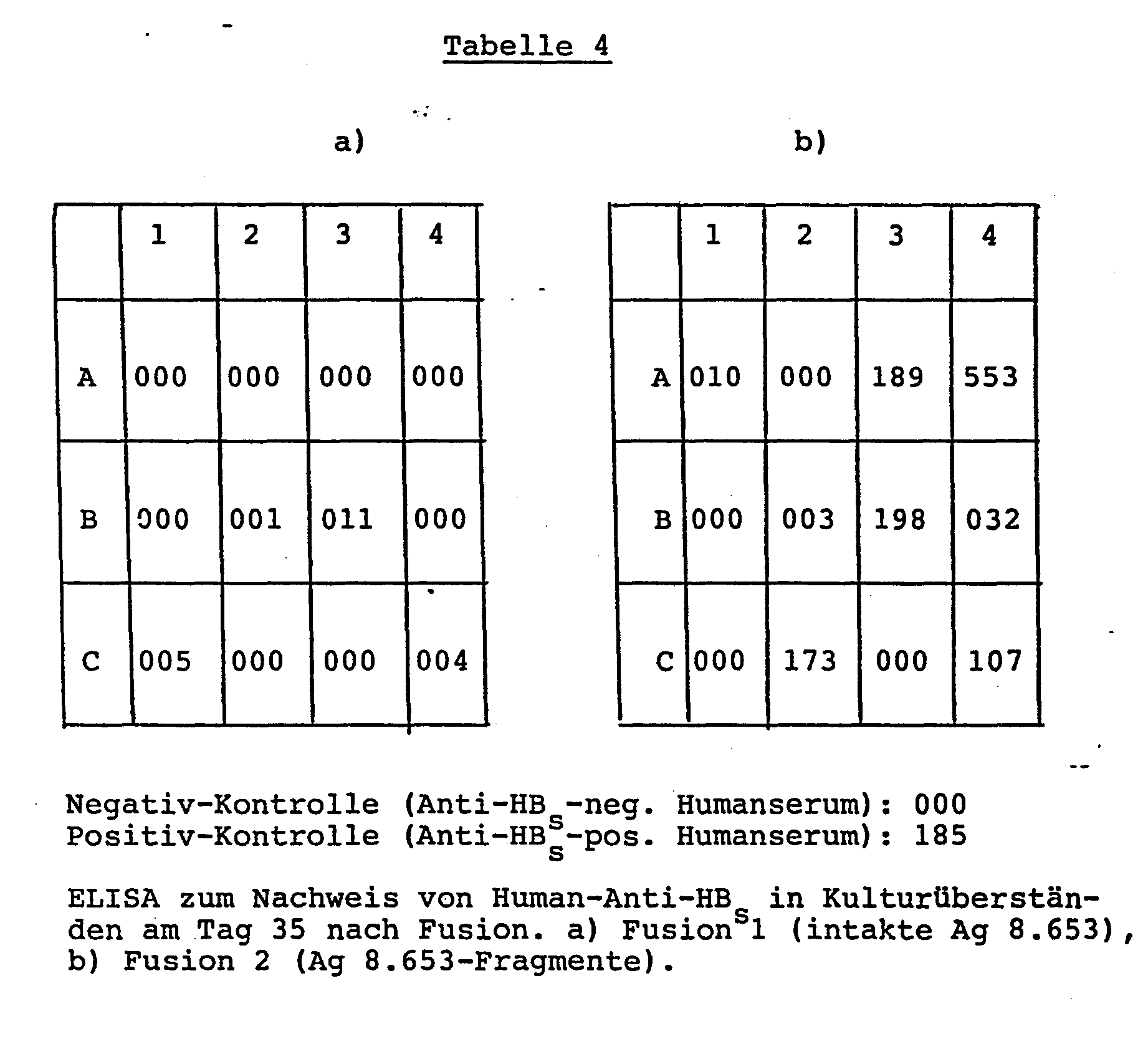
Fusion von HPBL mit Fragmenten aus der humanen Plasmacytom-Linie HS SULTANFusion of HPBL with fragments from the human plasmacytoma line HS SULTAN
HS Sultan wurde von der American Type Culture Collection (ATCC) unter dem Code CRL-1484 als kyropräserviertes Zellmaterial bezogen und nach ATCC-Vorschrift aufgetaut und in Kultur genommen.HS Sultan was obtained from the American Type Culture Collection (ATCC) under code CRL-1484 as a cell material that had been preserved in the cell, and was thawed in accordance with the ATCC specification and put into culture.
HPBL wurden wie in Beispiel 4 beschrieben vom gleichen Spender aufgearbeitet und in vitro mit HBsi "geboostert".HPBL were worked up as described in Example 4 from the same donor and "boosted" in vitro with HBsi.
Fusion: 4 x 10 HS SULTAN-Zellen wurden mittels Glycerin-Lyse fragmentiert. Die Membran-Cytoplasma-Vesikelfraktion wurde bei 5500 g (40 Minuten) sedimentiert und ein Gemisch von ca. 4 x 10⁷ HS-SULTAN-Kernen und 4 x 10⁷ HPBL(B) daraufgeschichtet. PEG-Fusion erfolgte nach Beispiel 4. Aussaat des Fusionats auf 12 24er Costar-Tüpfel in RPMI 1640 + 20 % FKS + Pyruvat + Insulin (Novo 2 U/ml) + 1 % nicht-essentielle Aminosäuren (BM) + 1 % Methocel 1500 ohne HAT-Zusatz.Fusion: 4 x 10 HS SULTAN cells were fragmented using glycerol lysis. The membrane cytoplasmic vesicle fraction was sedimented at 5500 g (40 minutes) and a mixture of approx. 4 × 10 7 HS-SULTAN cores and 4 × 10 7 HPBL (B) was layered on top. PEG fusion was carried out according to Example 4. Sowing the fusionate on 12 24-wells of Costar in RPMI 1640 + 20% FCS + pyruvate + insulin (Novo 2 U / ml) + 1% non-essential amino acids (BM) + 1% Methocel 1500 without HAT addition.
Tag 14 nach Fusion: Wachstum von großen, nichtadhärenten, lymphoiden Zellkolonien.Day 14 after fusion: growth of large, non-adherent, lymphoid cell colonies.
T-Lymphozyten wurden mittels Standardverfahren (Rosettierung mit Schaferythrozyten, Ficoll-Gradientenzentrifugation) aus der Lymphozyten-Gesamtfraktion isoliert und sofort oder nach 3tägiger Kultur behandelt. Ehrlich-Aszites-Zellen (ATCC; CCL 77) wurden in DMEM mit 10 % Pferdeserum kultiviert und dienten als Spender der transformierenden Fragmente. Die Fragmentierung der EAZ wurde nach Jett et al. (J. Biol. Chem. 252, 2134-2142 (1977)) mittels Glycerin-Lyse ausgeführt. Die überwiegend Kernmaterial enthaltende Fraktion wurde durch Zentrifugation abgetrennt und verworfen. Die Mitochondrien-reiche cytoplasmatische Membran-Vesikelfraktion (CMV) wurde zur Transformierung verwandt. 5 x 10⁷ T-Lymphozyten wurden mit einem Überschuß an PHA-Lektin (Difco) beladen, mit der CMV-Fraktion aus EAZ vermischt und 20 Minuten bei Raumtemperatur inkubiert. Die Mischung wurde durch Zentrifugation sedimentiert, der flüssige überstand restlos entfernt und durch 1 ml einer 50%igen PEG-Lösung ersetzt. Nach lminütiger Einwirkungszeit wurde die PEG-Lösung durch Zugabe von RPMI-1640-Kulturmedium ausverdünnt und durch Zentrifugation von den Zellen abgetrennt. Die Zellen wurden in RPMI-Medium mit 20 % foetalem Kälberserum (FKS, BM) aufgenommen, aus 12 1-ml-Kulturtüpfel verteilt und bei 37 °C in 5%iger C02-Atmosphäre kultiviert. Die Charakterisierung der Zellen anhand von Oberflächenmerkmalen erfolgte nach Standardverfahren mittels Schafserythrozyten (E)-Rosettierung (KAPLAN, M.E. et al.; J. Immunol. Methods 5, 131 (1974)) und mittels Immunfluoreszenz unter Verwendung der beiden T-Zell-spezifischen Antikörper OKT-3 (Ortho; REINHERZ, E.L. et al.; J. Immunol. 123, 1312 (1979)) bzw. MAK 4 - 11 (RIEBER, P. et al.; Hybridoma 1, 59 (1981)) und fluoreszenzmarkiertem Anti-Human-Immunglobulin (Ig; Fa. Dako) zum Nachweis von B-Zell-typischem Membran-Ig.T-lymphocytes were isolated from the total lymphocyte fraction using standard methods (rosetting with sheep erythrocytes, Ficoll gradient centrifugation) and treated immediately or after 3 days of culture. Ehrlich ascites cells (ATCC; CCL 77) were cultivated in DMEM with 10% horse serum and served as donors of the transforming fragments. The fragmentation of the EAZ was carried out according to Jett et al. (J. Biol. Chem. 252, 2134-2142 (1977)) by means of glycerol lysis. The fraction mainly containing core material was separated by centrifugation and discarded. The mitochondrial-rich cytoplasmic membrane vesicle fraction (CMV) was used for the transformation. 5 x 10 7 T lymphocytes were loaded with an excess of PHA lectin (Difco), mixed with the C MV fraction from EAZ and incubated for 20 minutes at room temperature. The mixture was sedimented by centrifugation, the liquid supernatant was removed completely and replaced by 1 ml of a 50% PEG solution. After an exposure time of 1 minute, the PEG solution was diluted by adding RPMI-1640 culture medium and separated from the cells by centrifugation. The cells were in RPMI medium with 20% fetal Calf serum (FKS, BM) ingested, distributed from 12 1 ml culture spots and cultivated at 37 ° C in a 5% C0 2 atmosphere. The characterization of the cells on the basis of surface features was carried out according to standard methods using sheep erythrocyte (E) rosetting (KAPLAN, ME et al .; J. Immunol. Methods 5, 131 (1974)) and using immunofluorescence using the two T cell-specific antibodies OKT-3 (Ortho; REINHERZ, EL et al .; J. Immunol. 123, 1312 (1979)) or MAK 4 - 11 (RIEBER, P. et al .; Hybridoma 1, 59 (1981)) and fluorescence-labeled anti Human immunoglobulin (Ig; Dako) for the detection of B-cell-typical membrane Ig.
Innerhalb der ersten 14 Tage starb der Großteil der Zellen in den Kulturen ab. Ab Tag 21 wurden im Zelldebris Kolonien von kleinen bis mittelgroßen Zellen sichtbar, die sich kontinuierlich vermehrten. Wachstum von Kolonien wurden in allen 12 Tüpfeln gefunden, und zwar bis zu 50 Kolonien pro__ Tüpfel. Am Tag 30 wurden die Kolonien in größere Kulturgefäße überführt und weiter vermehrt. Die Zellen wurden anhand ihrer Oberflächen-Marker analysiert und folgende Ergebnisse erhalten:

Fragmente, die aus EAZ (permanente Mauszell-Linie) durch Glycerin-Lyse gewonnen werden können, erzeugen nach PEG-Fusion mit isolierten T-Lymphozyten permanent in Kultur wachsende Zellen, die aufgrund ihrer Oberflächenmerkmale eindeutig als T-Lymphozyten identifizierbar sind.Fragments that can be obtained from EAZ (permanent mouse cell line) by glycerol lysis, after PEG fusion with isolated T-lymphocytes, produce cells that grow permanently in culture and that can be clearly identified as T-lymphocytes due to their surface characteristics.
Alle Kulturgefäße wurden vor Einsaat von Zellen mit Gelatine beschichtet. Das Kulturmedium bestand aus einer 1:1-Mischung von RPMI-1640 und Medium 199 (BM) mit 20 % FKS. Menschliche Endothelzellen wurden nach Jaffe et al. (J. Clin. Invest. 52, 2745-2756 (1973)) mittels Collagenase-Lösung (Gibco) aus den Venen von frischen Nabelschnüren gewonnen und vor der Transformierung durch Anlage einer Primärkultur über ca. 14 Tage vermehrt. Für die Transformierung wurden die adhärent wachsenden Endothelzellen mittels Trypsin-EDTA-Lösung (BM) abgelöst und in Suspension, wie unter 6.1 beschrieben, mit der CMV-Fraktion aus EAZ fusioniert. Die so behandelten Zellen wurden in einer Zelldichte von 5 x 10⁵ pro 75 cm2-Kulturgefäß ausgesät und im C02-Inkubator kultiviert. Zur Kontrolle wurden jeweils ein Aliquot der Endothelzellen aus den Primärkulturen ohne CMV-Fraktion mit PEG-Lösung behandelt (Scheinfusion) und rekultiviert. Sobald die Zellen auf dem Boden des Kulturgefäßes einen lückenlosen Zellrasen ausgebildet hatten, wurden sie mittels Trypsin-EDTA-Lösung abgelöst und im Verhältnis 1:3 auf frische Kulturgefäße übertragen (= Passage).All culture vessels were coated with gelatin before sowing cells. The culture medium consisted of a 1: 1 mixture of RPMI-1640 and medium 199 (BM) with 20% FCS. Human endothelial cells were developed according to Jaffe et al. (J. Clin. Invest. 52, 2745-2756 (1973)) by means of collagenase solution (Gibco) obtained from the veins of fresh umbilical cords and multiplied by transformation of a primary culture for about 14 days before transformation. For the transformation, the adherently growing endothelial cells were detached using trypsin-EDTA solution (BM) and fused in suspension, as described under 6.1, with the CMV fraction from EAZ. The cells treated in this way were sown in a cell density of 5 × 10 5 per 75 cm 2 culture vessel and cultured in a CO 2 incubator. As a control, an aliquot of the endothelial cells from the primary cultures without CMV fraction was treated with PEG solution (mock fusion) and recultivated. As soon as the cells had formed a complete cell lawn on the bottom of the culture vessel, they were detached using trypsin-EDTA solution and transferred to fresh culture vessels in a ratio of 1: 3 (= passage).
Die mit CMV-Fraktion fusionierten Endothelzellen hafteten nach Einsaat mit einer Effizienz von 20 bis 30 % an und wuchsen innerhalb von zwei bis drei Tagen zu einem konfluenten Zellrasen aus. Die schein-fusionierten Zellen hafteten mit etwa der gleichen Effizienz an, vermehrten sich aber kaum und starben - ohne einen konfluenten Zellrasen auszubilden - innerhalb von ca. 21 Tagen vollständig ab. Gänzlich unbehandelte Endothelzellen vermehrten sich bis maximal in die 3. Passage, stellten dann das Wachstum ein, lösten sich unter Abkugelung vom Boden des Kulturgefäßes und lysierten. In 8 verschiedenen Ansätzen mit Endothelzellen aus insgesamt 18 verschiedenen Nabelschnüren wuchsen die CMV-behandelten Zellen problemlos überdie 10. Passage hinaus. Die transformierten Endothelzellen konnten ohne Verlust an Lebens- und Teilungsfähigkeit in flüssigen Stickstoff ein- und ausgelagert werden.The endothelial cells fused with the CMV fraction adhered with an efficiency of 20 to 30% after sowing and grew into a confluent cell lawn within two to three days. The sham-fused cells adhered with about the same efficiency, but hardly increased and died completely within 21 days - without forming a confluent cell lawn. Completely untreated endothelial cells proliferated up to the third passage, then stopped growing, detached from the bottom of the culture vessel and lysed. In 8 different approaches with endothelial cells from a total of 18 different umbilical cords, the CMV-treated cells easily grew beyond the 10th passage. The transformed endothelial cells could be stored and removed in liquid nitrogen without loss of viability and ability to divide.
Humane Umbilical-Endothelzellen konnten ohne transformierende Manipulation gemäß Erfindung nicht über die 3. Passage kultiviert werden, womit die in der Literatur mitgeteilten Erfahrungen (z. B. Grimbrone, M.A., in Progress in Haemostasis and Thrombosis, Vol. 3; Maciag, T. et al., J. Cell Biol. 91, 420-426 (1981)) bestätigt wurden. Durch Fusion mit der Mitochondrien-reichen CMV-Fraktion aus Ehrlich-Aszites-Zellen wurden die Kultureigenschaften der.Endothelzellen so verändert, daß sie problemlos über die kritische 3. Passage hinaus vermehrbar waren (und sich derzeit in der 23. Passage befinden, ohne "Ermüdungserscheinungen" zu zeigen).Human umbilical endothelial cells could not be cultured via the third passage without transforming manipulation according to the invention, with which the experiences reported in the literature (e.g. Grimbrone, MA, in Progress in Haemostasis and Thrombosis, Vol. 3; Maciag, T. et al., J. Cell Biol. 91, 420-426 (1981)). By fusion with the mitochondrial-rich CMV fraction from Ehrlich-Ascites cells, the cultural properties of the endothelial cells were changed in such a way that they could be easily expanded beyond the critical 3rd passage (and are currently in the 23rd passage without " To show signs of fatigue ").
Melanomzell-haltiges Gewebe wurde durch chirurgische Exzidierung einer Hüft-Lymphknoten-Metastase gewonnen, unter sterilen Bedingungen in kleine Würfel zerschnitten und bis zur Fusion in flüssigen Stickstoff eingelagert. CMV-Fraktionen aus EAZ wurden, wie unter 6.1 beschrieben, hergestellt.Tissue containing melanoma cells was obtained by surgical excision of a hip-lymph node metastasis, cut into small cubes under sterile conditions and stored in liquid nitrogen until fusion. CMV fractions from EAZ were prepared as described under 6.1.
Vor der Fusion wurde das Melanomzell-haltige Material aufgetaut und mittels Trypsin-Behandlung einer Einzelzell-Suspension hergestellt. Die Fraktion der großen, braun-rot pigmentierten Melanomzellen wurden durch Ficoll-Gradiehten-Zentrifugation von den Begleit-Lymphozyten befreit und, wie unter 6.1 beschrieben, mit CMV-Fraktion unter PEG-Einfluß fusioniert (ca. 4 x 10⁵ Melanomzellen mit CMV aus ca. 1 x 10⁶ EAZ). Zur Fusionskontrolle dienten 4 x 10⁵ Melanomzellen, die ohne CMV mit PEG behandelt würden. Die Zellen wurden nach der Fusion in einer Dichte von 1 x 10⁵ Zellen pro Tüpfel in RPMI 1640 + 20 % FKS ausgesät und bei 37°C in 5%iger CO2-Atmosphäre kultiviert.Before the fusion, the melanoma cell-containing material was thawed and prepared by trypsin treatment of a single cell suspension. The fraction of the large, brown-red pigmented melanoma cells was freed from the accompanying lymphocytes by Ficoll graduate centrifugation and, as described under 6.1, fused with CMV fraction under the influence of PEG (approx. 4 × 10 5 melanoma cells with CMV approx. 1 x 10 6 EAZ). For merger control served 4 x 10 5 melanoma cells that would be treated with PEG without CMV. After the fusion, the cells were sown at a density of 1 × 10 5 cells per well in RPMI 1640 + 20% FCS and cultivated at 37 ° C. in a 5% CO 2 atmosphere.
In allen 4 Teilkulturen der mit CMV fusionierten Melanomzellen wuchsen ab Tag 28 Kolonien von typisch pigmentierten Zellen in Form von semiadhärenten Zellhaufen, die sich kontinuierlich vergrößerten. In der Kontroll-Kultur der schein-fusionierten Zellen kam es zu keiner Zellvermehrung, vielmehr zeigten die Zellen ab Tag 8 eine zunehmende Granulierung, und am Tag 22 waren nur noch Zelltrümmer in der Kultur vorhanden.From day 28, colonies of typically pigmented cells in the form of semi-adherent cell clusters grew in all 4 subcultures of the melanoma cells fused with CMV, which continuously increased. In the control culture of the sham-fused cells, there was no cell proliferation; rather, the cells showed an increasing granulation from day 8 and only cell debris remained in the culture on day 22.
Humane Melanomzellen aus kryopräserviertem Metastasengewebe konnten durch CMV-Fusion zu in vitro Kultur- und vermehrungsfähigen Zellen transformiert werden. Scheinfusionierte Melanomzellen gleicher Herkunft starben dagegen bei sonst identischen Kulturbedingungen ab.Human melanoma cells from cryopreserved metastatic tissue could be transformed by CMV fusion into in vitro culture and reproductive cells. In contrast, sham-fused melanoma cells of the same origin died under otherwise identical culture conditions.
Einführung eines eukaryontischen DNA-Vektors in immortalisierte menschliche Endothel-ZellenIntroduction of a eukaryotic DNA vector into immortalized human endothelial cells
Plasmid Z-pBR 322/RchrßG-Δ425 B (Dierks, P. et al., Proc. Natl. Acad. Sci. USA 78, 1411-1415 (1981)) enthält ein 2.070 Basenpaare (Bp) Kaninchen-ß-globin-Genfragment.Plasmid Z-pBR 322 / RchrßG-Δ425 B (Dierks, P. et al., Proc. Natl. Acad. Sci. USA 78, 1411-1415 (1981)) contains a 2,070 base pair (bp) rabbit-β-globin Gene fragment.
Das Cla-Pvu I-Fragment von pBR 322 wurde durch ein 3.039 Bp Hpa I-Bam H 1-Fragment ersetzt, welches die gesamte Region der frühen Gene und den Anfang der Region der späten Gene von SV 40 enthält (Tooze, J. (1980), in "DNA Tumor Viruses", J. Tooze, ed., 2nd edition, Cold Spring Harbor Laboratory und B. Wieringa et el., Cetus-UCLA Symposium on Gene regulation 1982). Dieses Plasmid wird im folgenden als pBR 322 RßG SV 40 bezeichnet.The Cla-Pvu I fragment of pBR 322 was replaced with a 3,039 bp Hpa I-Bam H 1 fragment which contains the entire region of the early genes and the beginning of the region of the SV 40 late genes (Tooze, J. ( 1980), in "DNA Tumor Viruses", J. Tooze, ed., 2nd edition, Cold Spring Harbor Laboratory and B. Wieringa et el., Cetus-UCLA Symposium on Gene regulation 1982). This plasmid is referred to below as pBR 322 RßG SV 40.
Menschliche Endothelzellen wurden, wie im Beispiel 7 beschrieben, immortalisiert und wachsen gelassen. Je 106 Zellen wurden mit 6 µg pBR 322 RßG SV 40 und mit 4 µg mit Ultraschall behandelter Kalbsthymus DNA transfiziert unter Anwendung der Kalziumphosphatfällungsmethode (Graham, F.L. et al., Virology 52, 456-467 (1973) und Wigler, M. et al., Cell 14, 725-731 (1978)). 10 Stunden und 40 Stunden nach der Transfektion wurden Hirt-Uberstände hergestellt (Hirt, B., J. Mol. Biol. 26, 365-369 (1967)).Human endothelial cells were immortalized and grown as described in Example 7. 10 6 cells each were transfected with 6 µg pBR 322 RßG SV 40 and with 4 µg ultrasound-treated calf thymus DNA using the calcium phosphate precipitation method (Graham, FL et al., Virology 52, 456-467 (1973) and Wigler, M. et al., Cell 14: 725-731 (1978)). Shepherd supernatants were prepared 10 hours and 40 hours after the transfection (Hirt, B., J. Mol. Biol. 26, 365-369 (1967)).
Die Hirt-Uberstände wurden auf einem l%igen Agarosegel getrennt und nach der Methode von Southern (Southern, E.M., J. Mol. Biol. 98, 503-517 (1975)) auf Nitrocellulosepapier übertragen. Plasmid pBR 322 RßG SV 40 wurde mit 32P unter Anwendung der Nick-Translationsmethode - von Rigby, P.W. et al., J. Mol. Biol. 113, 237-251 (1977) markiert und mit der übertragenen DNA auf den Nitrocellulosefiltern hybridisiert wie beschrieben bei Maniatis, T. et al., Molecular Cloning, Cold Spring Harbor Laboratory (1982). Der Filter wurde auf Röntgenfilm bei -70°C belichtet.The shepherd supernatants were separated on a 1% agarose gel and transferred to nitrocellulose paper by the method of Southern (Southern, EM, J. Mol. Biol. 98, 503-517 (1975)). Plasmid pBR 322 RßG SV 40 was labeled with 32 P using the Nick translation method - by Rigby, PW et al., J. Mol. Biol. 113, 237-251 (1977) and hybridized with the transferred DNA on the nitrocellulose filters as described in Maniatis, T. et al., Molecular Cloning, Cold Spring Harbor Laboratory (1982). The filter was exposed on X-ray film at -70 ° C.
Menschliche Endothelzellen, die mit Plasmid pBR 322 RßG SV 40 transfiziert wurden, ergeben ein starkes Hybridisierungssignal, welches dem authentischen Plasmid entspricht hinsichtlich seiner Mobilität auf Agarosegel. Ein Vergleich der Signale der Hirt-Uberstände der Zellen 40 Stunden nach der Transfektion mit denen der Zellen 10 Stunden nach der Transfektion ergab eine vier- bis fünffache Steigerung der Intensität des Hybridisierungssignals.Human endothelial cells which were transfected with plasmid pBR 322 RßG SV 40 give a strong hybridization signal which corresponds to the authentic plasmid with regard to its mobility on agarose gel. A comparison of the signals of the shepherd supernatants of the cells 40 hours after the transfection with those of the cells 10 hours after the transfection showed a four to five-fold increase in the intensity of the hybridization signal.
Eine Hybridisierung an DNA-Spezies, die kleiner sind als das eingebrachte Plasmid wurde ebenfalls beobachtet. Diese Signale waren in nicht transfizierten immortalisierten menschlichen Endothel- zellen nicht feststellbar.Hybridization to DNA species smaller than the introduced plasmid was also observed. These Signals were not found in non-transfected immortalized human endothelial cells.
Die gleiche Intensitätsverteilung der Hybridisierungssignale wurde beobachtet, wenn HeLa-Zellen mit Plasmid pBR 322 RßG SV 40 transfiziert wurden.The same intensity distribution of the hybridization signals was observed when HeLa cells were transfected with plasmid pBR 322 RßG SV 40.
Die Tatsache, daß in den Hirt-Uberständen menschlicher immortalisierter Epithelzellen, die mit Plasmid pBR 322 RßG SV 40 transfiziert worden waren, eine DNA-Hybridisierung an 32P-markiertem eingebrachtem Plasmid festgestellt wurde, jedoch nicht bei nichttransfizierten Zellen, ist ein Beweis dafür, daß der Vektor in die eukaryontische Zelllinie eingeführt wurde. Die Beobachtung, daß das Hybridisierungssignal bei transfizierten Zellen 40 Stunden nach der Transfektion vier- bis fünffach stärkere Intensität aufwies als bei Zellen 10 Stunden nach der Transfektion, läßt die Schlußfolgerung zu, daß das transfizierte Plasmid in den Zellen repliziert wurde.The fact that in the shepherd supernatants of human immortalized epithelial cells transfected with plasmid pBR 322 RßG SV 40, DNA hybridization to 32 P-labeled plasmid was found, but not in untransfected cells, is evidence that that the vector was introduced into the eukaryotic cell line. The observation that the hybridization signal in transfected cells was four to five times more intense 40 hours after the transfection than in cells 10 hours after the transfection leads to the conclusion that the transfected plasmid has been replicated in the cells.
Als Nuklease S1, T4 Polynukleotidkinase und Kälberdarmphosphatase wurden die handelsüblichen Präparate von Boehringer Mannheim verwendet.The commercially available preparations from Boehringer Mannheim were used as nuclease S 1 , T 4 polynucleotide kinase and calf intestinal phosphatase.
Immortalisierte menschliche Epithelzellen wurden wie in 9.1.2 beschrieben, vermehrt und mit dem das Kaninchen-ß-globin-Gen enthaltenden Vektor transfiziert. 48 Stunden nach der Transfektion wurden 1 bis 2 x 10⁶ Zellen lysiert und die RNA nach der LiCI-Harnstoff-Methode (Auffray, C. at al., Eur. J. Biochem. 107, 303-314 (1980)) extrahiert.Immortalized human epithelial cells were expanded as described in 9.1.2 and transfected with the vector containing the rabbit β-globin gene. 48 hours after the transfection, 1 to 2 × 10 6 cells were lysed and the RNA was extracted according to the LiCI-urea method (Auffray, C. at al., Eur. J. Biochem. 107, 303-314 (1980)).
Herstellung einer Kaninchen-ß-globin spezifischen Hybridisierungssonde.Preparation of a rabbit-ß-globin specific hybridization probe.
Plasmid pBR 322 RßG SV 40 wurde mit Hae III abgebaut und das sich von +135 bis -75 erstreckende Fragment (Dierks, P. et al., Proc. Natl. Acad. Sci. USA 78, 1411-1415 (1981)) und Van Oyen, A. et al., Science 206, 337-344 (19 ) wurde auf einem 2%igen Agarosegel isoliert und durch DEAE-Cellulose-Chromatographie weiter gereinigt (Müller, W. et al., J. Mol. Biol. 124, 343-358 (1978)). Das Fragment wurde mit Kälberdarmphosphatase und 32P-dephosphoriliert und mit γ-32P-ATP und T4 Polynukleotidkinase markiert wie beschrieben in (Mantei, N. et al., Gene 10, 1-8 (1980)).Plasmid pBR 322 RßG SV 40 was digested with Hae III and the fragment extending from +135 to -75 (Dierks, P. et al., Proc. Natl. Acad. Sci. USA 78, 1411-1415 (1981)) and Van Oyen, A. et al., Science 206, 337-344 (19) was isolated on a 2% agarose gel and further purified by DEAE cellulose chromatography (Müller, W. et al., J. Mol. Biol. 124, 343-358 (1978)). The fragment was treated with calf intestinal phosphatase and 32 P-dephosphorilated and labeled with γ- 32 P-ATP and T4 polynucleotide kinase as described in (Mantei, N. et al., Gene 10, 1-8 (1980)).
Das mit der Kinase behandelte Fragment wurde zusammen mit 10 µg E. coli t-RNA (Boehringer Mannheim) durch Äthanol gefällt und in 100 µl 0,4 M/l NaCl, 1 mM/1 EDTA, 40 mM/1 Pipes-Puffer, pH 6,4 und 80 % Formamid gelöst (Mantei, N. et al., Nature 281, 40-46 (1979). Die cellulare RNA (= 25 µg) wurde vakuumgetrocknet, in 10 µl Sonde (0,01 pMol, 30.000 cpm) in Puffer, wie oben beschrieben gelöst, denaturiert, in Glaskapillare eingeschmolzen und 16 Stunden bei 48°C hybridisiert. In einem Kontrollversuch wurde die Sonde mit Kaninchen-ß-globin m-RNA (Miles) hybridisiert. Die Probe wurde mit 100 µl 0,2 M/1 NaCl, 50 mM/1 Natriumacetatpuffer pH- 4,5, 1 mM/l ZnSO4 und 0,5 % Glycerin verdünnt und mit 50 Einheiten Nuklease S1 60 Minuten bei 30°C inkubiert (Weaver, R. et al., Nucl. Acid Res. 7, 1175-1193 (1979)).The fragment treated with the kinase was precipitated together with 10 μg E. coli t-RNA (Boehringer Mannheim) through ethanol and in 100 μl 0.4 M / l NaCl, 1 mM / 1 EDTA, 40 mM / 1 pipes buffer, pH 6.4 and 80% formamide dissolved (Mantei, N. et al., Nature 281, 40-46 (1979). The cellular RNA (= 25 μg) was vacuum-dried in 10 μl probe (0.01 pmol, 30,000 cpm) in buffer, as described above, denatured, melted in a glass capillary and hybridized for 16 hours at 48 ° C. In a control experiment, the probe was hybridized with rabbit-ß-globin m-RNA (Miles) 0.2 M / 1 NaCl, 50 mM / 1 sodium acetate buffer pH - 4.5, 1 mM / l ZnSO 4 and 0.5% glycerol diluted and incubated with 50 units of nuclease S 1 for 60 minutes at 30 ° C (Weaver, R . et al., Nucl. Acid Res. 7, 1175-1193 (1979)).
Die Proben wurden mit Phenol behandelt, zusammen mit 10 µg E. coli t-RNA durch Äthanol gefällt, mit 80 %igem Äthanol gewaschen (20 Minuten bei -70°C), vakuumgetrocknet, in 5 µl Färbelösung gelöst (0,05 % Bromphenolblau, 0,05 % Xylolxyanol, 1 mM/1 EDTA, 90 % V/V Formamid), 2 Minuten in einem siedenden Wasserbad erhitzt und der Elektrophorese auf 5 % Polyacrylamidgel (89 mM/1 Trisbase, 89 mM/1 Borsäure, 1 mM/l EDTA und 7 M/1 Harnstoff) unterworfen. Das Gel wurde mit Röntgenfilm der Firma Fuji und einem Verstärkerschirm bei -70°C autoradiographiert.The samples were treated with phenol, precipitated with ethanol together with 10 μg of E. coli t-RNA, washed with 80% ethanol (20 minutes at -70 ° C.), vacuum dried, dissolved in 5 µl staining solution (0.05% bromophenol blue, 0.05% xylene xyanol, 1 mM / 1 EDTA, 90% v / v formamide), heated in a boiling water bath for 2 minutes and electrophoresed on 5% polyacrylamide gel (89 mM / 1 Trisbase, 89 mM / 1 boric acid, 1 mM / l EDTA and 7 M / 1 urea). The gel was autoradiographed with X-ray film from Fuji and an intensifying screen at -70 ° C.
Die Größe der geschützten Fragmente konnte durch Vergleich mit Größenmarkern (32P-markiertes pBR 322 x Hinf 1 und pBR 322 x Hae III) beurteilt werden. Nicht transfizierte Zellen ergaben keinerlei globinspezifische Hybridisierungssignale (ausgenommen renaturierte Hybridisierungssonde = 210 Bp). Aus den transfizierten immortalisierten menschlichen Epithelzellen extrahierte RNA schützte zwei Fragmente, deren Größe mit 80 bis 90 Bp bestimmt wurde. Derartige Transkripte wurden auch durch Grosveld et al., Nature 295, 120-126 (1982) und Weidle, U. et al., Nature (1981) gefunden und werden dem Vorhandensein einer internen cryptischen Spleißstelle auf dem Kaninchen-ß-globin-Gen zugeschrieben. Transcripte, die von der cap-Stelle des Kaninchen-ß-globin-Gens stammen, konnten nicht festgestellt werden. In einem Vergleichsversuch wurden HeLa-Zellen mit Plasmid pBR 322 RßG SV 40 transfiziert. Korrekt iniziierte Transcripte (135 Bp geschützt) sowie Transcripte, die auf das Vorhandensein einer internen cryptischen Pleißstelle zurückgeführt werden (Grosveld, G. et al., Nature 295, - 120-126 (1982) und Weidle, U. et al., Nature (1983) wurden durch S1-Kartierung festgestellt.The size of the protected fragments could be assessed by comparison with size markers (32P-labeled pBR 322 x Hinf 1 and pBR 322 x Hae III). Untransfected cells gave no globin-specific hybridization signals (except for renatured hybridization probe = 210 bp). RNA extracted from the transfected immortalized human epithelial cells protected two fragments, the size of which was determined to be 80 to 90 bp. Such transcripts were also found by Grosveld et al., Nature 295, 120-126 (1982) and Weidle, U. et al., Nature (1981) and are based on the presence of an internal cryptic splice site on the rabbit β-globin gene attributed to. No transcripts from the cap site of the rabbit-ß-globin gene could be found. In a ver In the same experiment, HeLa cells were transfected with plasmid pBR 322 RßG SV 40. Correctly initiated transcripts (135 bp protected) as well as transcripts that are attributed to the presence of an internal cryptic splicing site (Grosveld, G. et al., Nature 295, - 120-126 (1982) and Weidle, U. et al., Nature (1983) were determined by S 1 mapping.
Die Tatsache, daß Kaninchen-ß-globin spezifische Transcripte in immortalisierten menschlichen Epithelzellen feststellbar sind, welche mit einem von SV 40 stammenden eukaryotischen Vektor, der das Kaninchen-ß-globin-Gen enthält, transfiziert wurden, zeigt, daß sich diese Zellinie als Wirtsystem für die Expression von wieder eingeführten geklonten Genen eignet.The fact that rabbit-ß-globin-specific transcripts are detectable in immortalized human epithelial cells which have been transfected with a SV 40-derived eukaryotic vector which contains the rabbit-ß-globin gene shows that this cell line is the host system suitable for the expression of re-introduced cloned genes.
Claims (8)
Priority Applications (1)
| Application Number | Priority Date | Filing Date | Title |
|---|---|---|---|
| AT83104243T ATE23361T1 (en) | 1982-05-04 | 1983-04-29 | PROCESSES FOR THE PRODUCTION OF PERMANENT ANIMAL AND HUMAN CELL LINES AND THEIR USE. |
Applications Claiming Priority (4)
| Application Number | Priority Date | Filing Date | Title |
|---|---|---|---|
| DE3216650 | 1982-05-04 | ||
| DE3216650 | 1982-05-04 | ||
| DE19823245665 DE3245665A1 (en) | 1982-05-04 | 1982-12-09 | METHOD FOR PRODUCING PERMANENT ANIMAL AND HUMAN CELL LINES AND THE USE THEREOF |
| DE3245665 | 1982-12-09 |
Publications (3)
| Publication Number | Publication Date |
|---|---|
| EP0093436A2 true EP0093436A2 (en) | 1983-11-09 |
| EP0093436A3 EP0093436A3 (en) | 1984-05-02 |
| EP0093436B1 EP0093436B1 (en) | 1986-11-05 |
Family
ID=25801552
Family Applications (1)
| Application Number | Title | Priority Date | Filing Date |
|---|---|---|---|
| EP83104243A Expired EP0093436B1 (en) | 1982-05-04 | 1983-04-29 | Process for preparing permanent animal and human cell lines, and their use |
Country Status (18)
| Country | Link |
|---|---|
| US (1) | US5114847A (en) |
| EP (1) | EP0093436B1 (en) |
| JP (1) | JPH0753110B2 (en) |
| AU (1) | AU538828B2 (en) |
| CA (1) | CA1201396A (en) |
| DD (1) | DD210708A5 (en) |
| DE (2) | DE3245665A1 (en) |
| DK (1) | DK199083A (en) |
| ES (1) | ES8402346A1 (en) |
| FI (1) | FI85282C (en) |
| GR (1) | GR78532B (en) |
| HU (1) | HU196456B (en) |
| IE (1) | IE54881B1 (en) |
| IL (1) | IL68515A (en) |
| LV (1) | LV11042B (en) |
| PT (1) | PT76629B (en) |
| SU (1) | SU1424744A3 (en) |
| YU (1) | YU94883A (en) |
Cited By (8)
| Publication number | Priority date | Publication date | Assignee | Title |
|---|---|---|---|---|
| DE3342736A1 (en) * | 1983-11-25 | 1985-06-05 | Boehringer Mannheim Gmbh, 6800 Mannheim | HYBRIDOMA ANTIBODY |
| WO1985003946A1 (en) * | 1984-03-07 | 1985-09-12 | Centre National De La Recherche Scientifique | Hybrid cells producing a determined polypeptide |
| DE3420926A1 (en) * | 1984-06-05 | 1985-12-05 | Boehringer Mannheim Gmbh, 6800 Mannheim | METHOD AND REAGENT FOR THE DIFFERENTIATED DETERMINATION OF ISOENZYMS OF ALKALINE PHOSPHATASE AND MONOCLONAL ANTIBODIES SUITABLE FOR THIS |
| WO1986007379A1 (en) * | 1985-06-11 | 1986-12-18 | The Upjohn Company | Transfer of male sterility in carrots |
| EP0239102A2 (en) * | 1986-03-28 | 1987-09-30 | Tsuji, Kimiyoshi | Process for the formation of human-human hybridoma |
| EP0292965A2 (en) * | 1987-05-27 | 1988-11-30 | Cetus Oncology Corporation | Human cell line and triomas, antibodes, and transformants derived therefrom |
| US5112748A (en) * | 1985-05-02 | 1992-05-12 | Institut Pasteur And Centre National De La Recherche Scientifique | Hybrid cells producing an antigen characteristic of the hepatitis b virus obtained from hepatocytes and established monkey cells, a process for obtaining these hybrid cells and their application to the production of the aforesaid antigen |
| US5126253A (en) * | 1983-01-20 | 1992-06-30 | Suntory Limited | Cell lines for use in the preparation of hybridoma cells |
Families Citing this family (10)
| Publication number | Priority date | Publication date | Assignee | Title |
|---|---|---|---|---|
| DE3525926A1 (en) | 1985-07-19 | 1987-01-29 | Boehringer Mannheim Gmbh | METHOD AND REAGENT FOR THE SPECIFIC DETERMINATION OF PANCREAS ALPHA AMYLASE |
| DE3627326A1 (en) * | 1986-08-12 | 1988-02-18 | Boehringer Mannheim Gmbh | IMMORTALIZATION THROUGH DNS TRANSMISSION |
| US5175004A (en) * | 1988-12-27 | 1992-12-29 | Matsumura Kenneth N | Propagatable, new combinant cells for cellular replacement therapy |
| US6515009B1 (en) | 1991-09-27 | 2003-02-04 | Neorx Corporation | Therapeutic inhibitor of vascular smooth muscle cells |
| US5811447A (en) | 1993-01-28 | 1998-09-22 | Neorx Corporation | Therapeutic inhibitor of vascular smooth muscle cells |
| US6491938B2 (en) | 1993-05-13 | 2002-12-10 | Neorx Corporation | Therapeutic inhibitor of vascular smooth muscle cells |
| US5981568A (en) | 1993-01-28 | 1999-11-09 | Neorx Corporation | Therapeutic inhibitor of vascular smooth muscle cells |
| US5358844A (en) * | 1993-02-18 | 1994-10-25 | Brigham And Women's Hospital, Inc. | Preservation of blood platelets |
| JP3096737B1 (en) * | 1999-07-19 | 2000-10-10 | 工業技術院長 | Human and animal cell culture |
| WO2023069575A1 (en) * | 2021-10-21 | 2023-04-27 | Parow Entheobiosciences Llc | Method for producing 5-methoxy-n,n-dimethyltryptamine (5-meo-dmt) in vitro |
Family Cites Families (7)
| Publication number | Priority date | Publication date | Assignee | Title |
|---|---|---|---|---|
| GB1594097A (en) * | 1976-12-16 | 1981-07-30 | Int Inst Of Differentiation | Production of specific immune nucleic acids cell dialysates and antibodies |
| US4394448A (en) * | 1978-02-24 | 1983-07-19 | Szoka Jr Francis C | Method of inserting DNA into living cells |
| US4172124A (en) * | 1978-04-28 | 1979-10-23 | The Wistar Institute | Method of producing tumor antibodies |
| JPS5852634B2 (en) * | 1980-12-05 | 1983-11-24 | 株式会社林原生物化学研究所 | Production method of urokinase |
| ZA824218B (en) * | 1981-06-29 | 1983-04-27 | Cetus Corp | Plasmid for producing human insulin |
| US4465769A (en) * | 1981-12-11 | 1984-08-14 | The United States Of America As Represented By The Department Of Health And Human Services | Non-transformed thymidine kinaseless cell line and its use for testing tumorigenic potential of genes |
| US4464465A (en) * | 1982-04-05 | 1984-08-07 | Genetic Systems Corporation | Cell-driven viral transfer in eukaryotes |
-
1982
- 1982-12-09 DE DE19823245665 patent/DE3245665A1/en not_active Withdrawn
-
1983
- 1983-04-20 GR GR71148A patent/GR78532B/el unknown
- 1983-04-25 IE IE945/83A patent/IE54881B1/en not_active IP Right Cessation
- 1983-04-26 YU YU00948/83A patent/YU94883A/en unknown
- 1983-04-28 IL IL68515A patent/IL68515A/en not_active IP Right Cessation
- 1983-04-29 EP EP83104243A patent/EP0093436B1/en not_active Expired
- 1983-04-29 DE DE8383104243T patent/DE3367412D1/en not_active Expired
- 1983-05-02 DD DD83250509A patent/DD210708A5/en not_active IP Right Cessation
- 1983-05-02 AU AU14141/83A patent/AU538828B2/en not_active Ceased
- 1983-05-03 PT PT76629A patent/PT76629B/en not_active IP Right Cessation
- 1983-05-03 SU SU833594155A patent/SU1424744A3/en active
- 1983-05-03 CA CA000427343A patent/CA1201396A/en not_active Expired
- 1983-05-03 HU HU831514A patent/HU196456B/en not_active IP Right Cessation
- 1983-05-03 FI FI831509A patent/FI85282C/en not_active IP Right Cessation
- 1983-05-04 ES ES522086A patent/ES8402346A1/en not_active Expired
- 1983-05-04 DK DK199083A patent/DK199083A/en not_active Application Discontinuation
-
1985
- 1985-08-23 JP JP60184369A patent/JPH0753110B2/en not_active Expired - Lifetime
-
1990
- 1990-09-25 US US07/590,602 patent/US5114847A/en not_active Expired - Fee Related
-
1993
- 1993-06-02 LV LVP-93-462A patent/LV11042B/en unknown
Non-Patent Citations (12)
| Title |
|---|
| A Dictionary of Immunology, Blackwell Scientific publications; Herbert W.J. and P.C. Wilkinson, 1977 2nd edition, page 97 * |
| BIOLOGICAL ABSTRACTS, Band 66, Nr. 5, 1. September 1978, Seite 2666, Zusammenfassung Nr. 27083, Philadelphia, US * |
| Cell and Tissue culture- John Paul, 5th Edition, Churchill Livingstone, pp. 82-83 * |
| CHEMICAL ABSTRACTS, Band 82, Nr. 23, 9. Juni 1975, Seite 252, Nr. 151810h, Columbus, Ohio, US * |
| CHEMICAL ABSTRACTS, Band 86, Nr. 17, 25. April 1977, Seite 360, Nr. 118951q, Columbus, Ohio, US * |
| CHEMICAL ABSTRACTS, Band 86, Nr. 21, 23. Mai 1977, Seite 202, Nr. 152366n, Columbus, Ohio, US * |
| Immunology, John Wiley and Sons, pages 43 and 47 * |
| MEDICAL LABORATORY SCIENCES, Band 36, 1979, Seiten 329-338, The Institute of Medical Laboratory Sciences * |
| Molecular Biology of "The Cell", Garland Publishing, Inc., Bruce Alberts et al., p. 163 * |
| Nucleic Acids Research, Vol. 5, no. 7, July 1978, p.2313-19 * |
| Proceedings National Academy of Sciences USA, Vol. 78, N. 3, March 1981, pp. 1411-15 * |
| Sciences, Vol. 206, 19 October 1979, P. 337-344 * |
Cited By (14)
| Publication number | Priority date | Publication date | Assignee | Title |
|---|---|---|---|---|
| US5126253A (en) * | 1983-01-20 | 1992-06-30 | Suntory Limited | Cell lines for use in the preparation of hybridoma cells |
| DE3342736A1 (en) * | 1983-11-25 | 1985-06-05 | Boehringer Mannheim Gmbh, 6800 Mannheim | HYBRIDOMA ANTIBODY |
| EP0150309A2 (en) * | 1983-11-25 | 1985-08-07 | Roche Diagnostics GmbH | Hybridoma antibody |
| EP0150309A3 (en) * | 1983-11-25 | 1985-08-21 | Boehringer Mannheim Gmbh | Hybridoma antibody |
| WO1985003946A1 (en) * | 1984-03-07 | 1985-09-12 | Centre National De La Recherche Scientifique | Hybrid cells producing a determined polypeptide |
| EP0158546A1 (en) * | 1984-03-07 | 1985-10-16 | Centre National De La Recherche Scientifique (Cnrs) | Specific polypeptide producing hybrid cell lines obtained from primary cells which are spontaneously capable of expressing said polypeptide or an equivalent polypeptide, process for obtaining the hybrid cells and their use in the production of said polypeptide |
| WO1985005690A1 (en) * | 1984-06-05 | 1985-12-19 | Boehringer Mannheim Gmbh | Method and reactant for the differential determination of isoenzymes of the alkaline phosphatase and monoclonal antibody therefor |
| DE3420926A1 (en) * | 1984-06-05 | 1985-12-05 | Boehringer Mannheim Gmbh, 6800 Mannheim | METHOD AND REAGENT FOR THE DIFFERENTIATED DETERMINATION OF ISOENZYMS OF ALKALINE PHOSPHATASE AND MONOCLONAL ANTIBODIES SUITABLE FOR THIS |
| US5112748A (en) * | 1985-05-02 | 1992-05-12 | Institut Pasteur And Centre National De La Recherche Scientifique | Hybrid cells producing an antigen characteristic of the hepatitis b virus obtained from hepatocytes and established monkey cells, a process for obtaining these hybrid cells and their application to the production of the aforesaid antigen |
| WO1986007379A1 (en) * | 1985-06-11 | 1986-12-18 | The Upjohn Company | Transfer of male sterility in carrots |
| EP0239102A2 (en) * | 1986-03-28 | 1987-09-30 | Tsuji, Kimiyoshi | Process for the formation of human-human hybridoma |
| EP0239102A3 (en) * | 1986-03-28 | 1989-07-12 | Tsuji, Kimiyoshi | Process for the formation of human-human hybridoma |
| EP0292965A2 (en) * | 1987-05-27 | 1988-11-30 | Cetus Oncology Corporation | Human cell line and triomas, antibodes, and transformants derived therefrom |
| EP0292965A3 (en) * | 1987-05-27 | 1990-01-03 | Cetus Oncology Corporation | Human cell line and triomas, antibodes, and transformants derived therefrom |
Also Published As
| Publication number | Publication date |
|---|---|
| US5114847A (en) | 1992-05-19 |
| EP0093436B1 (en) | 1986-11-05 |
| YU94883A (en) | 1988-10-31 |
| EP0093436A3 (en) | 1984-05-02 |
| FI831509L (en) | 1983-11-05 |
| IL68515A0 (en) | 1983-07-31 |
| DK199083D0 (en) | 1983-05-04 |
| IE54881B1 (en) | 1990-03-14 |
| LV11042B (en) | 1996-06-20 |
| LV11042A (en) | 1996-02-20 |
| DE3367412D1 (en) | 1986-12-11 |
| PT76629A (en) | 1983-06-01 |
| HU196456B (en) | 1988-11-28 |
| DD210708A5 (en) | 1984-06-20 |
| AU538828B2 (en) | 1984-08-30 |
| JPS61205486A (en) | 1986-09-11 |
| CA1201396A (en) | 1986-03-04 |
| FI831509A0 (en) | 1983-05-03 |
| ES522086A0 (en) | 1984-02-01 |
| IL68515A (en) | 1988-11-15 |
| IE830945L (en) | 1983-11-04 |
| SU1424744A3 (en) | 1988-09-15 |
| AU1414183A (en) | 1983-11-10 |
| DK199083A (en) | 1983-11-05 |
| GR78532B (en) | 1984-09-27 |
| JPH0753110B2 (en) | 1995-06-07 |
| PT76629B (en) | 1985-12-03 |
| ES8402346A1 (en) | 1984-02-01 |
| FI85282B (en) | 1991-12-13 |
| DE3245665A1 (en) | 1983-11-10 |
| FI85282C (en) | 1992-03-25 |
Similar Documents
| Publication | Publication Date | Title |
|---|---|---|
| EP0093436B1 (en) | Process for preparing permanent animal and human cell lines, and their use | |
| DE3301249C2 (en) | ||
| JPH02227096A (en) | Antibody and preparation thereof | |
| AT400577B (en) | METHOD FOR PRODUCING A MONOCLONAL ANTIBODY AND METHOD FOR DETECTING MALIGNAL CELLS | |
| DE2942780A1 (en) | EUKARYOTIC CELLS, EUKARYOTIC PROTOPLASTICS AND MULTI-CELL EUKARYOTIC LIVING ORGANISMS CONTAINING DNA IN LIPID VESICLES, METHODS FOR THE PRODUCTION OF GENE PRODUCTS, FOR IMMEDIATING, AND DEFECTED BREADING | |
| DE3249567C2 (en) | Hybrid cell line, process for its production and its use | |
| DE10050935A1 (en) | Use of CD28-specific monoclonal antibodies to stimulate blood cells that do not carry CD28 | |
| CH683526A5 (en) | Cell line for the production of antibody-producing hybridomas, methods for producing a human cell line, and antibody-producing hybridoma. | |
| AT395017B (en) | METHOD FOR PRODUCING A NEW LYMPHOKIN (LK 2) AND USE OF LK 2 FOR PRODUCING A PHARMACEUTICAL PREPARATION | |
| DE3239863C2 (en) | Process for the preparation of a subculturable lymphokine-producing human T-cell hybridoma | |
| DE3413339A1 (en) | ANTIBODIES AGAINST MUSHROOMS OF THE GENE CANDIDA | |
| EP0256512B1 (en) | Immortalization by dna transfer | |
| DE19647426C1 (en) | Human colon carcinoma cell lines with stable HSP72 expression | |
| EP0090343A2 (en) | Hybrid cell, process for preparaing it and its use | |
| Buxbaum et al. | Analysis of Thy-1 variants of murine lymphoma cells | |
| DD241917A5 (en) | Use of permanently culturable human or animal cell line | |
| JPS6239999B2 (en) | ||
| EP0157271B1 (en) | Hybrid cell line, process for preparing it and its use | |
| US6013518A (en) | Fused cell line and method of obtaining the same | |
| EP0576862A2 (en) | DNA for immortalisation of human or animal cells | |
| DD243713A1 (en) | PROCESS FOR OBTAINING MONOCLONAL ANTIBODIES AGAINST RAT PERITONEAL MAST CELLS | |
| EP0547076A1 (en) | Cd58-specific monoclonal antibodies and their use | |
| CH670099A5 (en) | New hybridoma cell lines - obtd. by fusing mouse-human hybrid cells with human cells | |
| DE3616324A1 (en) | MONOCLONAL ANTIBODIES | |
| JPS59187795A (en) | Antiaspergillus fungus antibody |
Legal Events
| Date | Code | Title | Description |
|---|---|---|---|
| PUAI | Public reference made under article 153(3) epc to a published international application that has entered the european phase |
Free format text: ORIGINAL CODE: 0009012 |
|
| AK | Designated contracting states |
Designated state(s): AT BE CH DE FR GB IT LI LU NL SE |
|
| PUAL | Search report despatched |
Free format text: ORIGINAL CODE: 0009013 |
|
| AK | Designated contracting states |
Designated state(s): AT BE CH DE FR GB IT LI LU NL SE |
|
| 17P | Request for examination filed |
Effective date: 19840529 |
|
| GRAA | (expected) grant |
Free format text: ORIGINAL CODE: 0009210 |
|
| AK | Designated contracting states |
Kind code of ref document: B1 Designated state(s): AT BE CH DE FR GB IT LI LU NL SE |
|
| REF | Corresponds to: |
Ref document number: 23361 Country of ref document: AT Date of ref document: 19861115 Kind code of ref document: T |
|
| ITF | It: translation for a ep patent filed |
Owner name: ING. C. GREGORJ S.P.A. |
|
| REF | Corresponds to: |
Ref document number: 3367412 Country of ref document: DE Date of ref document: 19861211 |
|
| ET | Fr: translation filed | ||
| ITTA | It: last paid annual fee | ||
| PLBE | No opposition filed within time limit |
Free format text: ORIGINAL CODE: 0009261 |
|
| STAA | Information on the status of an ep patent application or granted ep patent |
Free format text: STATUS: NO OPPOSITION FILED WITHIN TIME LIMIT |
|
| 26N | No opposition filed | ||
| EPTA | Lu: last paid annual fee | ||
| EAL | Se: european patent in force in sweden |
Ref document number: 83104243.7 |
|
| PGFP | Annual fee paid to national office [announced via postgrant information from national office to epo] |
Ref country code: SE Payment date: 20000406 Year of fee payment: 18 |
|
| PGFP | Annual fee paid to national office [announced via postgrant information from national office to epo] |
Ref country code: FR Payment date: 20000411 Year of fee payment: 18 |
|
| PGFP | Annual fee paid to national office [announced via postgrant information from national office to epo] |
Ref country code: AT Payment date: 20000412 Year of fee payment: 18 |
|
| PGFP | Annual fee paid to national office [announced via postgrant information from national office to epo] |
Ref country code: LU Payment date: 20000419 Year of fee payment: 18 |
|
| PGFP | Annual fee paid to national office [announced via postgrant information from national office to epo] |
Ref country code: GB Payment date: 20000426 Year of fee payment: 18 |
|
| PGFP | Annual fee paid to national office [announced via postgrant information from national office to epo] |
Ref country code: DE Payment date: 20000427 Year of fee payment: 18 Ref country code: CH Payment date: 20000427 Year of fee payment: 18 |
|
| PGFP | Annual fee paid to national office [announced via postgrant information from national office to epo] |
Ref country code: NL Payment date: 20000428 Year of fee payment: 18 |
|
| PGFP | Annual fee paid to national office [announced via postgrant information from national office to epo] |
Ref country code: BE Payment date: 20000622 Year of fee payment: 18 |
|
| PG25 | Lapsed in a contracting state [announced via postgrant information from national office to epo] |
Ref country code: LU Free format text: LAPSE BECAUSE OF NON-PAYMENT OF DUE FEES Effective date: 20010429 Ref country code: GB Free format text: LAPSE BECAUSE OF NON-PAYMENT OF DUE FEES Effective date: 20010429 Ref country code: AT Free format text: LAPSE BECAUSE OF NON-PAYMENT OF DUE FEES Effective date: 20010429 |
|
| PG25 | Lapsed in a contracting state [announced via postgrant information from national office to epo] |
Ref country code: SE Free format text: LAPSE BECAUSE OF NON-PAYMENT OF DUE FEES Effective date: 20010430 Ref country code: FR Free format text: THE PATENT HAS BEEN ANNULLED BY A DECISION OF A NATIONAL AUTHORITY Effective date: 20010430 Ref country code: BE Free format text: LAPSE BECAUSE OF NON-PAYMENT OF DUE FEES Effective date: 20010430 |
|
| PG25 | Lapsed in a contracting state [announced via postgrant information from national office to epo] |
Ref country code: LI Free format text: LAPSE BECAUSE OF NON-PAYMENT OF DUE FEES Effective date: 20010528 Ref country code: CH Free format text: LAPSE BECAUSE OF NON-PAYMENT OF DUE FEES Effective date: 20010528 |
|
| BERE | Be: lapsed |
Owner name: BOEHRINGER MANNHEIM G.M.B.H. Effective date: 20010430 |
|
| PG25 | Lapsed in a contracting state [announced via postgrant information from national office to epo] |
Ref country code: NL Free format text: LAPSE BECAUSE OF NON-PAYMENT OF DUE FEES Effective date: 20011101 |
|
| EUG | Se: european patent has lapsed |
Ref document number: 83104243.7 |
|
| REG | Reference to a national code |
Ref country code: CH Ref legal event code: PL |
|
| GBPC | Gb: european patent ceased through non-payment of renewal fee |
Effective date: 20010429 |
|
| NLV4 | Nl: lapsed or anulled due to non-payment of the annual fee |
Effective date: 20011101 |
|
| PG25 | Lapsed in a contracting state [announced via postgrant information from national office to epo] |
Ref country code: DE Free format text: LAPSE BECAUSE OF NON-PAYMENT OF DUE FEES Effective date: 20020201 |
|
| REG | Reference to a national code |
Ref country code: FR Ref legal event code: ST |