EP0091005A1 - Method for detecting the presence of GD3 ganglioside - Google Patents
Method for detecting the presence of GD3 ganglioside Download PDFInfo
- Publication number
- EP0091005A1 EP0091005A1 EP83102865A EP83102865A EP0091005A1 EP 0091005 A1 EP0091005 A1 EP 0091005A1 EP 83102865 A EP83102865 A EP 83102865A EP 83102865 A EP83102865 A EP 83102865A EP 0091005 A1 EP0091005 A1 EP 0091005A1
- Authority
- EP
- European Patent Office
- Prior art keywords
- cells
- abr
- melanoma
- gangliosides
- ganglioside
- Prior art date
- Legal status (The legal status is an assumption and is not a legal conclusion. Google has not performed a legal analysis and makes no representation as to the accuracy of the status listed.)
- Granted
Links
Images


Classifications
-
- G—PHYSICS
- G01—MEASURING; TESTING
- G01N—INVESTIGATING OR ANALYSING MATERIALS BY DETERMINING THEIR CHEMICAL OR PHYSICAL PROPERTIES
- G01N33/00—Investigating or analysing materials by specific methods not covered by groups G01N1/00 - G01N31/00
- G01N33/48—Biological material, e.g. blood, urine; Haemocytometers
- G01N33/50—Chemical analysis of biological material, e.g. blood, urine; Testing involving biospecific ligand binding methods; Immunological testing
- G01N33/53—Immunoassay; Biospecific binding assay; Materials therefor
- G01N33/574—Immunoassay; Biospecific binding assay; Materials therefor for cancer
- G01N33/57407—Specifically defined cancers
- G01N33/5743—Specifically defined cancers of skin, e.g. melanoma
-
- G—PHYSICS
- G01—MEASURING; TESTING
- G01N—INVESTIGATING OR ANALYSING MATERIALS BY DETERMINING THEIR CHEMICAL OR PHYSICAL PROPERTIES
- G01N33/00—Investigating or analysing materials by specific methods not covered by groups G01N1/00 - G01N31/00
- G01N33/48—Biological material, e.g. blood, urine; Haemocytometers
- G01N33/50—Chemical analysis of biological material, e.g. blood, urine; Testing involving biospecific ligand binding methods; Immunological testing
- G01N33/53—Immunoassay; Biospecific binding assay; Materials therefor
- G01N33/5308—Immunoassay; Biospecific binding assay; Materials therefor for analytes not provided for elsewhere, e.g. nucleic acids, uric acid, worms, mites
-
- Y—GENERAL TAGGING OF NEW TECHNOLOGICAL DEVELOPMENTS; GENERAL TAGGING OF CROSS-SECTIONAL TECHNOLOGIES SPANNING OVER SEVERAL SECTIONS OF THE IPC; TECHNICAL SUBJECTS COVERED BY FORMER USPC CROSS-REFERENCE ART COLLECTIONS [XRACs] AND DIGESTS
- Y10—TECHNICAL SUBJECTS COVERED BY FORMER USPC
- Y10S—TECHNICAL SUBJECTS COVERED BY FORMER USPC CROSS-REFERENCE ART COLLECTIONS [XRACs] AND DIGESTS
- Y10S436/00—Chemistry: analytical and immunological testing
- Y10S436/804—Radioisotope, e.g. radioimmunoassay
-
- Y—GENERAL TAGGING OF NEW TECHNOLOGICAL DEVELOPMENTS; GENERAL TAGGING OF CROSS-SECTIONAL TECHNOLOGIES SPANNING OVER SEVERAL SECTIONS OF THE IPC; TECHNICAL SUBJECTS COVERED BY FORMER USPC CROSS-REFERENCE ART COLLECTIONS [XRACs] AND DIGESTS
- Y10—TECHNICAL SUBJECTS COVERED BY FORMER USPC
- Y10S—TECHNICAL SUBJECTS COVERED BY FORMER USPC CROSS-REFERENCE ART COLLECTIONS [XRACs] AND DIGESTS
- Y10S436/00—Chemistry: analytical and immunological testing
- Y10S436/811—Test for named disease, body condition or organ function
- Y10S436/813—Cancer
-
- Y—GENERAL TAGGING OF NEW TECHNOLOGICAL DEVELOPMENTS; GENERAL TAGGING OF CROSS-SECTIONAL TECHNOLOGIES SPANNING OVER SEVERAL SECTIONS OF THE IPC; TECHNICAL SUBJECTS COVERED BY FORMER USPC CROSS-REFERENCE ART COLLECTIONS [XRACs] AND DIGESTS
- Y10—TECHNICAL SUBJECTS COVERED BY FORMER USPC
- Y10S—TECHNICAL SUBJECTS COVERED BY FORMER USPC CROSS-REFERENCE ART COLLECTIONS [XRACs] AND DIGESTS
- Y10S436/00—Chemistry: analytical and immunological testing
- Y10S436/815—Test for named compound or class of compounds
-
- Y—GENERAL TAGGING OF NEW TECHNOLOGICAL DEVELOPMENTS; GENERAL TAGGING OF CROSS-SECTIONAL TECHNOLOGIES SPANNING OVER SEVERAL SECTIONS OF THE IPC; TECHNICAL SUBJECTS COVERED BY FORMER USPC CROSS-REFERENCE ART COLLECTIONS [XRACs] AND DIGESTS
- Y10—TECHNICAL SUBJECTS COVERED BY FORMER USPC
- Y10S—TECHNICAL SUBJECTS COVERED BY FORMER USPC CROSS-REFERENCE ART COLLECTIONS [XRACs] AND DIGESTS
- Y10S436/00—Chemistry: analytical and immunological testing
- Y10S436/828—Protein A
Definitions
- the antigen detected by Ab R 24 is identified herein as G D3- a previously characterized disialoganglioside.
- melanomas have high levels of G D3 .
- these antibodies are useful in determining whether a tissue sample is a melanoma or not. This is particularly important for characterizing lesions.
- These antibodies can also be used in determining concentrations of G D3 in serum, plasma, urine or other body fluids. This may aid in the early diagnosis of melanoma and possibly of other disorders where there are elevated glycolipid levels.
- Mouse monoclonal antibody AbR 24 (Dippold et al., Proc. Natl. Acad. Sci. 77: 6114-6118, 1980) has a high degree of specificity for human melanoma cells when tested on viable cultured cells using the PA-MHA serological assay.
- the antigen detected by this antibody has been isolated from melanoma cells and shown to be G D3 ganglioside by compositional and partial structural analysis and by comparison with authentic G D3 by thin layer chromatography (TLC).
- TLC thin layer chromatography
- GMIA glycolipid-mediated immune adherence
- AbR 24 , AbC 5 , AbI 12 , and AbN 9 have been previously described (1).
- AbR 24 and AbC 5 are I gG3 antibodies and AbI 12 and AbN 9 are IgG2b and IgGl antibodies, respectively.
- G D3 was obtained from Dr. Y-T. Li, Tulane University, New York (5).
- G M3 and G M2 were obtained from Drs. S. Kundu and D. M. Marcus, Baylor University, Houston, TX.
- G M1 , G D1a , G T1 were purchased from Supelco, Inc., Bellefonte, PA. Lactosylceramide was purchased from Glycolipid Biochemical Co., Birmingham, Al.
- Reactivity of AbR 24 and AbC 5 with sell surface antigens of melanoma cells was determined with cultured cells growing in the wells of Microtest plates (Falcon 3034) using a red cell rosetting method (3) in which indicator cells are human 0 red cells (RBC) to which Staphylococcus aureus protein A is conjugated (PA-MHA).
- indicator cells are human 0 red cells (RBC) to which Staphylococcus aureus protein A is conjugated (PA-MHA).
- AbI 12 and AbN 9 were assayed using a modification of this method in which rabbit anti-mouse Ig-conjugated indicator cells were used (IgG-MHA).
- HBSS Hank's balanced salt solution
- Vibro cholerae neuraminidase Cal- biochem
- B-galactosidase Sigma, Type VII
- Glycolipids were isolated initially by a modification of the method of Saito and Hakomori (6), and separated into neutral and acidic fractions by DEAE-Sephadex chromatography (7). Acidic glycolipids (gangliosides) were subsequently isolated directly from chloroform-methanol (C-M) extracts by DEAE-Sephadex chromatography and alkaline hydrolysis (7). Briefly, cells were homogenized in C-M (2 : 1) and after filtration were reextracted with C-M (1 : 1). After evaporating and redissolving the extract in C-M (1 : 2), it is filtered, evaporated and dialyzed against distilled ice water for 24 hours in the cold.
- C-M chloroform-methanol
- Lipid-bound sialic acid in cell pellets was determined on C:M (2 : 1 and 1 : 2) extracts after hydrolysis in 0.1N HCl at 80° for 1 hour as described by Warren (10). Sugars were analysed after methanolysis (methanolic 1.0 N HCl at 100° for 16 hours) as their 0-trifluoroacetates (11); N-acetylneuraminic acid was identified by the same procedure after methanolysis in 1.0.N HCl at 80° for 1.0 hour.
- Glycolipids (6 ⁇ g sialic acid) were dissolved, aliquoted into tubes (10 x 75 mm) and dried in a dessicator with P 2 0 5 in vacuo. To each tube, 200 ⁇ l of PBS was added, the sides of the tube scraped and the solutions sonicated for 15 min at 50° C. After transfer to a larger tube, 0.8 ml of PBS was added. The glycolipid solution was added slowly in a dropwise fashion to a 2 % suspension of human O-RBC in PBS. After 1 hour at 37° C, with one mixing after 30 minutes, the cells were washed twice with PBS (12 ml each wash). Reactivity was tested by mixing a suspension of the treated RBC and appropriately diluted AbR 24 (50 ⁇ l each) in microtiter plates. After 1-2 hours at 4° C, the agglutination reactions were scored visually.
- Glycolipids (6 ⁇ g sialic acid), dissolved in C-M (1 : 2), were aliquoted into tubes (6 x 50 mm) and dried as in the previous assay.
- AbR 24 (1:2 x 10 ) was added (30 ⁇ l) and the tubes were vor- texed, incubated for 30 minutes at room temperature, and then for 30 minutes at 4° C. Tubes were centrifuged for 20 minutes at 2000 rpm and the supernatants removed and serially diluted. These samples were immediately transferred to form- aldehydefixed SK-MEL 28 target cells. (The formaldehyde fixation was carried out by treating cells growing in the wells of microtest plates (Falcon 3034) with 0.33 % HCHO in PBS. This treatment does not alter reactivity with AbR 24 and provides a store of readily available source of target cells). Antibody reactivity was detected with the PA-MHA assay. Unabsorbed antibody served as a positive control.
- a solution of glycolipids in 95 % ethanol was added to the wells of microtest plates (Falcon 3034; 10 ⁇ l per well) and the plates were dried in a dessicator in vacuo with P 2 0 5 for 45 minutes. Approximately 100 ng of lipid-bound sialic acid was found to be the optimal amount for efficient adsorption and maximal reactivity with antibody.
- Wells were then washed three times with PBS-2 % GG-free FBS (10 ml/wash), and the plates blotted with gauze. Diluted antibody (in PBS with 5 % GG-free FBS) was added to the wells' and incubated for 45 minutes at room temperature.
- SK-MEL-28 melanoma cells After treatment with neuraminidase (Vibrio cholerae), SK-MEL-28 melanoma cells no longer reacted with AbR 24 in PA-MHA assays (Table I). Reactivity with AbC S (an antibody with a serological specificity similar to that of AbR 24 (1)) was also lost. Reactivity with AbN 9 and AbI12 which recognize serologically unrelated determinants on glycoproteins of SK-MEL-28 was unaffected by neuraminidase. Enzyme-treated cells did not show non-specific reactivity with either Protein A-or with anti-mouse Ig-indicator cells.
- Glycolipids were isolated from cultured melanoma cells (SK-MEL 28) by chloroform-methanol (C-M) extraction and Florisil chromatography of their acetates as described by Saito and Hakomori (5) and the glycolipid preparation was fractionated into neutral and acidic components by DEAE-Sephadex chromatography. Inhibitory activity against AbR 24 antibody (assayed with PA-MHA) was found to reside entirely in the acidic glycolipid fractions.
- the isolated AbR 24 -reactive glycolipid was identified as G D3 [NANA(2 ⁇ 8)NANA(2 ⁇ 3)Ga1ß(1 ⁇ 4)Glc-ceramide] by the following criteria:
- G D3 Distribution of G D3 in -melanoma and non-melanoma cell lines, and in normal and malignant tissues
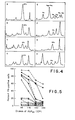
- Total ganglioside fractions were prepared from a large variety of tumor cell lines, fresh tumors and normal tissues. When these extracts were fractionated by TLC and the gangliosides detected using the resorcinol reagent, it became evident that melanomas have a characteristic pattern of gangliosides. In all the melanoma cell lines examined, glycolipids comigrating with G D3 and G M3 were prominent acidic glycolipids, with G D3 being the major component in many of these cell lines ( Figure 3 and Figure 4). G D3 was also a prominent ganglioside in extracts of mouse eye and bovine choroid. With the exception of MOLT-4 (a T-cell line), none of the other cells or tissues had G D3 as the major component.
- MOLT-4 a T-cell line
- Extracts of fresh melanoma tumors gave ganglioside patterns resembling SK-MEL-28, with G D3 and G M3 p redomi- nating (Figure 3). Most melanoma cell lines gave this simlified pattern, but some showed a more complex profile in which higher gangliosides were detected in appreciable amounts ( Figures 3 and 4). G D3 constituted 18-63 % of the total ganglioside fraction in the melanoma cell lines examined ( Figure 4). Most melanoma cell lines and specimens had values in the 30 - 50 % range.
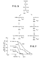
- G D3 could be detected in extracts of melanoma cell lines and melanoma tissue, but not in other sources (Table II). More sensitive assays (inhibition of PA-MHA and GMIA methods) showed that G D3 was detectable in a wider range of cells (bovine choroid, mouse eye, fetal and adult human lung, RAJI B-cell line, MOLT-4 T-cell line, RT-4 bladder cancer cell line and AJ astro- cytoma cell line). A typical inhibition experiment is presented in Figure 5 and the data are summarized in Table II.
- Mouse monoclonal antibody R24 which shows a high degree of serological specificity for cell surface antigens of melanoma cells, recognizes a disialoganglioside - G D3 .
- Past studies have shown that antibodies to gangliosides have been difficult to raise (15).'This may have to do with the fact that most gangliosides are constituents of the species being immunized and also, because in situ sialidase activity may destroy ganglioside immunogenicity (16). In this regard, it might be significant that the mouse from which AbR 24 was developed had been extensively immunized over a period of six months with melanoma cells (SK-MEL-28) having a very high G D3 content.
- G D3 is a prominent ganglioside in cultured melanoma cells and in melanoma tissue. When compared with other cells, melanoma cells also posses relatively high total ganglioside levels. As shown by others, G D3 is present in small amounts in most mammalian tissues, but it is a major ganglioside in the retina, where it comprises between 30 - 40 % of the gangliosides (19). In adult human brain. G D3 represents about 8 - 10 % of the total ganglioside conterit (19).
- G D3 may be higher in fetal brain, considering that in fetal rat brain (15 - 17 days gestation) G D3 represents about 50 % of the total ganglioside content, falling rapidly to about 10 % by day 20 (20).
- Portoukalian and coworkers (21) have also reported that G D3' identified by TLC and carbohydrate analysis, is a major constituent of melanomas. They showed that the proportion of GD3 varied from 31.0 % to 57.2 % of the ganglioside fraction in the four different melanoma specimens examined. From these results, as well as our own analysis, one can conclude that G D3 ganglioside is a prominent component of malignant melanoma.
- T-cell line MOLT-4 showed a similar profile, and this may be another example of antigens shared by T-cells and cells of neuroectodermal origin e. g. Thy-1 (25).
- Gangliosides derived from bovine choroid and mouse eye had more complex patterns, with G D3 being only one of three or four prominent components.
- G D3 ganglioside is by no means restricted to melanoma cells - it is ubiquitous. Yet using direct serological assays for cell surface antigens, only melanomas, choroidal melanocytes, and astrocytomas were reactive with AbR 24 (1). Even using sensitive absorption tests, only normal brain of other cells and tissues tested absorbed AbR 24 . A number of explanations for the apparent discrepancy between the serological finding and the biochemical data presented here can be suggested. First, it is possible that G D3 is not a cell surface constituent of most non-melanoma cells.
- G D3 is a biosynthetic precursor of other gangliosides (Figur 9) and would therefore be located mainly within the cell, probably in the Golgi apparatus where the glycosyl transferases responsible for glycolipid synthesis are found (26, 27). As our biochemical studies were carried out on whole cell or tissues the results are certainly compatible with this explanation. Another possibility is that G D3 is present at the cell surface of R 24 -negative cells but is not available for reaction with antibody. This phenomenon has been found with other cell membrane glycolipids e. g. globoside is a major glycolipid of erythrocyte membrane but erythrocytes react only weakly with anti-globoside antibody (28).
- G D3 is not expressed on the surface of most non-melanoma cells in amounts that are detectable by the serological tests used. It is important to note that the cell types which reacted with AbR 24 in both direct and absorption tests have both a high lipid-bound sialic acid content and have G D3 as a prominent ganglioside.
- melanomas have high levels of B-N-acetylgalactosaminidase which would result in increased degradation of G M2 and G D2 or that melanomas have elevated levels of certain sialytrans- ferases, resulting in increased synthesis of G D3 and G M3 .
- melanoma patients have increased serum sialyltransferase levels (29).
- Enzyme levels in tumor tissue have not yet been studied, although the fact that the glycoproteins of human melanoma cell lines have increased sialylation as compared to the glycoproteins of other cell types (30) suggests increased activity of this enzyme in melanoma.
- TLC thin layer chromatography
- MHA mixed hemagglutination assay
- C-M Chloroform-methanol
- FBS fetal bovine serum
- NANA N-acetylneuraminic acid
- Ga1 D-galactose
- Glc D-glucose
- GalNAc N-acetyl-D-galactosamine
- Cer ceramide
- G M1 BGal 1 - 3 GalNAC 1 ⁇ 4 BGal (3 ⁇ 2 NANA) 1 ⁇ 4 G1c-Cer
- G M3 NANA 2 ⁇ 3 BGal 1 ⁇ 4 Glc-Cer
- G D3 NANA 2 ⁇ 8 NANA 2 ⁇ 3 BGal 1 ⁇ 4 Glc-Cer
- G M2 BGalNAc 1-4 BGal (3 ⁇ 2 NANA)
- G lc
Abstract
- (a) passive hemmaglutination
- (b) antibody inhibition
- (c) glycolipid-mediated immune adherence
- (d) a method using 125|-Protein A to detect AbR24 reacting with GD3 on thin layer chromatograms.
Description
- We have previously described a mouse IgG3 monoclonal antibody (AbR24) with a high degree of serological specificity for cultured human melanoma cells (1). All melanoma cell lines examined and two astrocytomas were positive for the heatstable cell surface antigen detected by this antibody. Although choroidal melanocytes and brain had low levels of the antigen, a wide variety of other cells and tissues were unreactive. Three other monoclonal antibodies (Abs C51 I24, and K9), having a similar restricted specificity, were derived from the same fusion. These antibodies showed the same strong reactivity with melanomas and lack of reactivity with epithelial cells, but had a slightly wider specificity range in that they also reacted weakly with MOLT-4 (a T-cell line), leukocytes and some fetal tissues.
- The antigen detected by Ab R24 is identified herein as GD3- a previously characterized disialoganglioside. In comparison with other cells and tissues, melanomas have high levels of GD3. Thus, these antibodies are useful in determining whether a tissue sample is a melanoma or not. This is particularly important for characterizing lesions. These antibodies can also be used in determining concentrations of GD3 in serum, plasma, urine or other body fluids. This may aid in the early diagnosis of melanoma and possibly of other disorders where there are elevated glycolipid levels.
- Mouse monoclonal antibody AbR24 (Dippold et al., Proc. Natl. Acad. Sci. 77: 6114-6118, 1980) has a high degree of specificity for human melanoma cells when tested on viable cultured cells using the PA-MHA serological assay. The antigen detected by this antibody has been isolated from melanoma cells and shown to be GD3 ganglioside by compositional and partial structural analysis and by comparison with authentic GD3 by thin layer chromatography (TLC). AbR24 reacts with authentic GD3' but not with any other ganglioside tested. Using TLC and reactivity with AbR24, a wide range of cells and tissues was examined for the presence of GD3. A new serological assay, termed glycolipid-mediated immune adherence (GMIA), was devised for assaying the reactivity of AbR24 with gangliosides. Melanomas (cultured cells or tumor tissue) were shown to have GD3 and GM3 as major gangliosides. Other cells and tissues examined also contained GD3' but usually only in low amounts. Melanomas (and MOLT-4, a T-cell line) were characterized by a simplified ganglioside profile with GD3 and GM3 as major components. The apparent discrepancy between the ubiquitous presence of GD3 and the serological specificity of AbR24 for melanoma cells can be explained in terms of localization and concentration of GD3 in different cells.
-
- Figure 1. Time course for the reexpression of AbR24- reactive antigen on SK-MEL-28 cells after neuraminidase treatment. Assay: PA-MHA.
- Figure 2. Localization of AbR24-reactive glycolipid on thin layer chromatography using glycolipid-mediated immune adherence (GMIA) assay. Acidic glycolipids from SK-MEL-28 cells were separated by TLC in
solvent 1. Silica gel bands (1 cm wide) were scraped from the plate, extracted with C:M (1.2) and assayed for antigens by GMIA as described in the text. - Figure 3. Thin layer chromatography of acidic glycolipid fractions from a number of cell lines and tissues. Lane 1: GM3; 2: GD3; 3: GM1; 4: SK-Mel-28 melanoma cell line; 5: AbR24-reactive antigen isolated from SK-MEL-28; 6: SK-MEL-37 melanoma cell line; 7: SK-MEL-64 melanoma cell line; 8: MeWo; 9: SK-MEL-13 melanoma cell line; 10: melanoma (surgical specimen); 11: MOLT-4 T-cell line; 12: Mouse eye; 13: SK-RC-7 renal cancer cell line; 14: adult human brain. Gangliosides were separated in
solvent 1 and visualized with resorcinol-HCl. - Figure 4. Densiometric tracings of thin layer chromatograms of gangliosides from melanomas and other cells. A: SK-MEL-28 melanoma cell line; B: SK-MEL-37 melanoma cell line; C: SK-MEL-13 melanoma cell line; D: melanoma (surgical specimen); E: adult human brain; F: Raji B-cell line; G: MOLT-4 cell line; H: SK-RC-7 renal cell line. The amount of GD3, as % of total ganglioside fraction, was calculated from the areas of the peaks and is indicated in each panel.
- Figure 5. Inhibition of reactivity of AbR24 with SK-MEL-28 melanoma cells by acidic glycolipid fractions from a variety of cell lines and tissues. Assay: PA-MHA.: AbR24;
 : adult human spleen; • : adult human liver; 0:.teleost eye; ■ : SK-RC-7 renal cancer cell line; ▲ : adult human brain;
 : MeWo melanoma cell line; ◆ : SK-MEL-29 melanoma cell line; V : SK-MEL-37 melanoma cell line; V : mouse eye; □ : MOLT-4 T-cell line; Δ : melanoma (surgical specimen);
 : SK-MEL-28 melanoma cell line.
- Figure 6. Glycolipid-mediated immune adherence (GMIA) assay using AbR24. Well A: AbR24-reactive glycolipid isolated from SK-MEL-28 melanoma cell line; well B: GD3 ganglioside; well C: no ganglioside; well D: GM2 and GM3 ganglio- side mixture. Antibody: AbR24 (1 : 1000).
- Figure 7. Detection of GD3 ganglioside by AbR24 in GMIA assays. AbR24 dilutions are indicated in the figure.
- Figure 8. Detection of GD3 ganglioside on TLC plates by reactivity with AbR24 and 125I-Protein A. Right side: gangliosides visualized with resorcinol-HC1 reagent;left side: gangliosides reacting with AbR24 and 125I-Protein A. Lane 1: AbR24-reactive ganglioside; Lane 2: gangliosides extracted from adult human brain.
Solvent 2. - Figure 9. Proposed pathways for the biosynthesis of gangliosides (modified after Yu and Ando (32)).
- For derivations and culture of melanoma and other cells see references 1 - 4. Normal and malignant tissue was obtained from surgical or postmortem specimens.
- Mouse monoclonal antibodies AbR24, AbC5, AbI12, and AbN9 have been previously described (1). AbR24 and AbC5 are IgG3 antibodies and AbI12 and AbN9 are IgG2b and IgGl antibodies, respectively.
- GD3 was obtained from Dr. Y-T. Li, Tulane University, New Orleans (5). GM3 and GM2 were obtained from Drs. S. Kundu and D. M. Marcus, Baylor University, Houston, TX. GM1, GD1a, GT1 were purchased from Supelco, Inc., Bellefonte, PA. Lactosylceramide was purchased from Glycolipid Biochemical Co., Birmingham, Al.
- Serological assays for melanoma cell surface antigens
- Reactivity of AbR24 and AbC5 with sell surface antigens of melanoma cells was determined with cultured cells growing in the wells of Microtest plates (Falcon 3034) using a red cell rosetting method (3) in which indicator cells are human 0 red cells (RBC) to which Staphylococcus aureus protein A is conjugated (PA-MHA). AbI12 and AbN9 were assayed using a modification of this method in which rabbit anti-mouse Ig-conjugated indicator cells were used (IgG-MHA).
- Melanoma cells growing as monolayers in microtest plates as described above were washed with Hank's balanced salt solution (HBSS, Microbiological Associates) and then treated with Vibro cholerae neuraminidase (Cal- biochem) or B-galactosidase (Sigma, Type VII) using 1 U/well in 10 µl of HBSS. After incubation for 1 hr at 370, the cells were washed four times with PBS - 2 % gamma globulin (GG)-free FBS and assayed for reactivity with antibody using the PA- or IgG-MHA assays.
- Glycolipids were isolated initially by a modification of the method of Saito and Hakomori (6), and separated into neutral and acidic fractions by DEAE-Sephadex chromatography (7). Acidic glycolipids (gangliosides) were subsequently isolated directly from chloroform-methanol (C-M) extracts by DEAE-Sephadex chromatography and alkaline hydrolysis (7). Briefly, cells were homogenized in C-M (2 : 1) and after filtration were reextracted with C-M (1 : 1). After evaporating and redissolving the extract in C-M (1 : 2), it is filtered, evaporated and dialyzed against distilled ice water for 24 hours in the cold. After dialysis, samples were evaporated, dissolved in C-M-H20 (30 : 60 : 8) and applied to a DEAE-Sephadex column (equilibrated with C-M-0.8M Na acetate); (30 : 60 : 8). After washing the column with C-M-H20 (30 : 60 : 8), acidic lipids were eluted with C-M-0.8M Na acetate (30 : 60 : 8), evaporated and dialyzed as before. The acidic fraction was then hydrolyzed with 0.1 N NaOH in methanol for 3 hours at 37° C, dialyzed against cold water (48 hours), evaporated, and dissolved in C-M (4 : 1). The solution was applied to a Biosil A column which had previously been washed with C-M (4 : 1). After eluting impurities with C-M (4 : 1), gangliosides were eluted with C-M (1 : 2).
- Silica gel plates (Rediplates, Fisher Scientific Co.) were activated by heating at 120° C for 1 hour. Solvents used for developing chromatograms were n-propanol-NH40H-
H 20, 60 : 9.5 : 11.5 (solvent 1) as in ref. 8 and chloroform: methanol: 2.5N NH40H, 60 : 40 : 9 (solvent 2). Once the solvent had migrated 12 cm from the origin, the plate was removed and air-dried for 12 - 15 minutes at 110 - 120° C, cooled to room temperature and sprayed with resorcinol -HCl (9). For preparative analysis, plates were dried at room temperature in an air flow hood for 2 - 3 hours. Bands were visualized with iodine vapor, outlined and silica gel scraped from the plate. The gel was then extracted twice with 40 ml of C-M-H20 (50 : 50 : 15), with a small amount of Dowex 50 (Na+). The suspension was centrifuged at 1000 rpm for 15 minutes and the solution filtered, evaporated, redissolved in C-M (4 : 1) and applied to a Biosil A column as described above. Impurities were eluted with C-M (4 : 1) and adsorbed gangliosides were then eluted with C-M (1 : 2). - Lipid-bound sialic acid in cell pellets was determined on C:M (2 : 1 and 1 : 2) extracts after hydrolysis in 0.1N HCl at 80° for 1 hour as described by Warren (10). Sugars were analysed after methanolysis (methanolic 1.0 N HCl at 100° for 16 hours) as their 0-trifluoroacetates (11); N-acetylneuraminic acid was identified by the same procedure after methanolysis in 1.0.N HCl at 80° for 1.0 hour.
- Glycolipids (6µg sialic acid) were dissolved, aliquoted into tubes (10 x 75 mm) and dried in a dessicator with
P 205 in vacuo. To each tube, 200 µl of PBS was added, the sides of the tube scraped and the solutions sonicated for 15 min at 50° C. After transfer to a larger tube, 0.8 ml of PBS was added. The glycolipid solution was added slowly in a dropwise fashion to a 2 % suspension of human O-RBC in PBS. After 1 hour at 37° C, with one mixing after 30 minutes, the cells were washed twice with PBS (12 ml each wash). Reactivity was tested by mixing a suspension of the treated RBC and appropriately diluted AbR24 (50 µl each) in microtiter plates. After 1-2 hours at 4° C, the agglutination reactions were scored visually. - Glycolipids (6µg sialic acid), dissolved in C-M (1 : 2), were aliquoted into tubes (6 x 50 mm) and dried as in the previous assay. AbR24 (1:2 x 10 ) was added (30 µl) and the tubes were vor- texed, incubated for 30 minutes at room temperature, and then for 30 minutes at 4° C. Tubes were centrifuged for 20 minutes at 2000 rpm and the supernatants removed and serially diluted. These samples were immediately transferred to form- aldehydefixed SK-MEL 28 target cells. (The formaldehyde fixation was carried out by treating cells growing in the wells of microtest plates (Falcon 3034) with 0.33 % HCHO in PBS. This treatment does not alter reactivity with AbR24 and provides a store of readily available source of target cells). Antibody reactivity was detected with the PA-MHA assay. Unabsorbed antibody served as a positive control.
- A solution of glycolipids in 95 % ethanol was added to the wells of microtest plates (Falcon 3034; 10 µl per well) and the plates were dried in a dessicator in vacuo with
P 205 for 45 minutes. Approximately 100 ng of lipid-bound sialic acid was found to be the optimal amount for efficient adsorption and maximal reactivity with antibody. Wells were then washed three times with PBS-2 % GG-free FBS (10 ml/wash), and the plates blotted with gauze. Diluted antibody (in PBS with 5 % GG-free FBS) was added to the wells' and incubated for 45 minutes at room temperature. Plates were blotted, washed four times with PBS-2 % GG-free FBS, and blotted again. Ten µl of a 0.2 % suspension of Protein A-conjugated O-RBC were added to the wells. The plates were incubated for 30 minutes at room temperature. After blotting, the plates were washed twice with PBS-2% GG-free FBS, blotted once again and read under the light microscope. Reactions were scored according to the proportion of the well which was covered by red cells. A test was read as negative when wells showed no adhering cells or only a thin ring of cells around the perimeter. - Serological reactivity of glycolipids separated by thin layer chromatography was tested using a modification of the method of Magnani et al. (13) in which 125I-Protein A was used to detect the bound antibody. After chromatography in
solvents - After treatment with neuraminidase (Vibrio cholerae), SK-MEL-28 melanoma cells no longer reacted with AbR24 in PA-MHA assays (Table I). Reactivity with AbCS (an antibody with a serological specificity similar to that of AbR24 (1)) was also lost. Reactivity with AbN9 and AbI12 which recognize serologically unrelated determinants on glycoproteins of SK-MEL-28 was unaffected by neuraminidase. Enzyme-treated cells did not show non-specific reactivity with either Protein A-or with anti-mouse Ig-indicator cells. B-Galactosidase had no detectable effect on the reactivity of SK-MEL-28 cells with AbR24 or AbC5 (Table I). These results show that sialic acid constitutes an important part of the antigenic determinant recognized by antibodies AbR24 and AbC S.
- Serological reactivity of AbR24 with SK-MEL-28 remained undetectable for 30 minutes after neuraminidase was removed and replaced with MEM-FBS. Continued incubation in this medium at 37° resulted in a partial return of AbR24 reactivity after 2 hours and complete recovery of serological reactivity after 22 hours (Figure 1).
- Glycolipids were isolated from cultured melanoma cells (SK-MEL 28) by chloroform-methanol (C-M) extraction and Florisil chromatography of their acetates as described by Saito and Hakomori (5) and the glycolipid preparation was fractionated into neutral and acidic components by DEAE-Sephadex chromatography. Inhibitory activity against AbR24 antibody (assayed with PA-MHA) was found to reside entirely in the acidic glycolipid fractions.
- In subsequent experiments, acidic glycolipids from SK-MEL-28 cells were isolated directly by fractionating the C-M extract on DEAE-Sephadex (6) and eliminating contaminating phospholipids by alkaline hydrolysis. Individual gangliosides in this mixture were isolated by preparative thin layer chromatography in solvent 1 (8). By scraping a series of silica gel bands from the plates and extracting the glycolipids, the antigenic activity was located in the major acidic glycolipid band which migrated just above GM1 and GDla (Figure 2). In the data presented in Figure 2, the antigenic activity of fractions was measured by the GMLA assay. Similar results were obtained by antibody inhibition tests using the PA-MHA assay with AbR24 and SK-MEL-28 target cells.
- The isolated AbR24-reactive glycolipid was identified as GD3 [NANA(2→8)NANA(2→3)Ga1ß(1→4)Glc-ceramide] by the following criteria:
- (I) carbohydrate analysis of the purified glycolipid showed that it contained glucose, galactose and N-acetylneuraminic acid in a ratio of 1.0 : 1.09 : 2.11 with only a trace (< 0.1) of hexosamine,
- (II) partial hydrolysis of the ganglioside with Vibrio cholerae neuraminidase (3 hours at 370 C) resulted in the formation of two components comigrating on thin layer chromatograms with GM3 and lactosylceramide,
- (III) the purified melanoma glycolipid comigrated with authentic GD3 in thin layer chromatography (Figure 3) and
- (IV) AbR24 reacted with authentic GD3, but not with any of the other standard gangliosides tested (see below).
- Total ganglioside fractions were prepared from a large variety of tumor cell lines, fresh tumors and normal tissues. When these extracts were fractionated by TLC and the gangliosides detected using the resorcinol reagent, it became evident that melanomas have a characteristic pattern of gangliosides. In all the melanoma cell lines examined, glycolipids comigrating with GD3 and GM3 were prominent acidic glycolipids, with GD3 being the major component in many of these cell lines (Figure 3 and Figure 4). GD3 was also a prominent ganglioside in extracts of mouse eye and bovine choroid. With the exception of MOLT-4 (a T-cell line), none of the other cells or tissues had GD3 as the major component. Extracts of fresh melanoma tumors gave ganglioside patterns resembling SK-MEL-28, with GD3 and GM3 predomi- nating (Figure 3). Most melanoma cell lines gave this simlified pattern, but some showed a more complex profile in which higher gangliosides were detected in appreciable amounts (Figures 3 and 4). GD3 constituted 18-63 % of the total ganglioside fraction in the melanoma cell lines examined (Figure 4). Most melanoma cell lines and specimens had values in the 30 - 50 % range. These values compared with 7 % in adult human brain, 9 % in a renal cancer cell line (SK-RC-7), 11 % in RAJI cells (a Burkitt's lymphoma) and 33 % in MOLT-4 cells (Figure 4). In terms of the serological reactivity of AbR24, it is important to note that melanomas, in addition to having higher proportions of GD3 in their glycolipid fraction, also have higher total ganglioside levels. This is evident from a determination of the levels of lipid-bound sialic acid in a number of cell lines. In melanomas the values ranged from 0.039 - 0.063 µ mole/0.1 ml cells (determined on 9 lines). RAJI, MOLT-4 and renal cancer cells (3 lines) had lipid-bound sialic acid values of 0.011 ± 0.003, 0.013 ± 0.006 and 0.025 - 0.029 4 mole/0.1 ml cells, respectively.
- (II) Detection of GD3 in cell lines and tissues using AbR24 antibody GD3 levels in a large variety of cells and tissues were estimated using R24 antibody. Four assay methods were used:
- (a) passive hemagglutination,
- (b) antibody inhibition,
- (c) a new method, GMIA, devised to combine the simplicity of the MHA method with the ability of glycolipids to adsorb to plastic and
- (d) a method using 125I-Protein A to detect AbR24 reacting with GD3 on TLC chromatograms. The sensitivity of the assays varies considerably; the passive hemagglutination assay is the least sensitive and the 125 I-PA method the most sensitive (Table II).
- Using the least sensitive detection method (passive hemagglutination), GD3 could be detected in extracts of melanoma cell lines and melanoma tissue, but not in other sources (Table II). More sensitive assays (inhibition of PA-MHA and GMIA methods) showed that GD3 was detectable in a wider range of cells (bovine choroid, mouse eye, fetal and adult human lung, RAJI B-cell line, MOLT-4 T-cell line, RT-4 bladder cancer cell line and AJ astro- cytoma cell line). A typical inhibition experiment is presented in Figure 5 and the data are summarized in Table II. Using the GMIA method it was found that wells coated with R24-reactive glycolipids from melanoma (Figure 6A) or authentic GD3 gave strongly positive reactions (Figure 6B); some quantitative data on this reaction are shown in Figure 7. Other purified glycolipids (GM1, GD1a' G M3 and GM2) were unreactive in this assay (Table II and Figure 6 D). AbR24 added alone was also unreactive (Figure 6C). Application of this method to acidic glycolipids extracted from other cells gave approximately the same results as inhibition assays (Table II). In contrast to the restricted distribution of GD3 determined by these methods, the 125I-Protein A method detected GD3 in all the cells and tissues examined (Table II). That the AbR24-reactive component detected in these tissues and cells was in fact GD3 was indicated by its co-migration with authentic GD3 (in two solvent systems), and by the finding that another protein A-binding monoclonal antibody (AbI12), detecting an unrelated glycoprotein specificity, was unreactive.
- Mouse monoclonal antibody R24, which shows a high degree of serological specificity for cell surface antigens of melanoma cells, recognizes a disialoganglioside - GD3. Past studies have shown that antibodies to gangliosides have been difficult to raise (15).'This may have to do with the fact that most gangliosides are constituents of the species being immunized and also, because in situ sialidase activity may destroy ganglioside immunogenicity (16). In this regard, it might be significant that the mouse from which AbR24 was developed had been extensively immunized over a period of six months with melanoma cells (SK-MEL-28) having a very high GD3 content. Two other monoclonal antibodies recognizing gangliosides have recently been described (17, 18). One reacts specifically with chicken neuronal cells and is directed against one of the higher gangliosides present in the GQ fraction (17); the second is directed against human colon carcinoma and recognizes an as yet uncharacterized monosialo- ganglioside (18).
- We have shown that GD3 is a prominent ganglioside in cultured melanoma cells and in melanoma tissue. When compared with other cells, melanoma cells also posses relatively high total ganglioside levels. As shown by others, GD3 is present in small amounts in most mammalian tissues, but it is a major ganglioside in the retina, where it comprises between 30 - 40 % of the gangliosides (19). In adult human brain. GD3 represents about 8 - 10 % of the total ganglioside conterit (19). Levels of GD3 may be higher in fetal brain, considering that in fetal rat brain (15 - 17 days gestation) GD3 represents about 50 % of the total ganglioside content, falling rapidly to about 10 % by day 20 (20). Portoukalian and coworkers (21) have also reported that GD3' identified by TLC and carbohydrate analysis, is a major constituent of melanomas. They showed that the proportion of GD3 varied from 31.0 % to 57.2 % of the ganglioside fraction in the four different melanoma specimens examined. From these results, as well as our own analysis, one can conclude that GD3 ganglioside is a prominent component of malignant melanoma. Whether normal melanocytes have high levels of GD3 is at present unclear. Normal choroidal melanocytes show weak reactivity with AbR24 in direct serological tests (titer 1 : 100) as compared to the strong reactivity of melanoma cells (titer of 1 : 5 x 10⁴ - 105) (1). With the recent development of a method for culturing skin melanocytes (22), it will now be possible to make a direct comparison of the GD3 content of melanocytes and melanomas. Although a precise biological funtion for GD3 remains to be determined, it has been suggested that GD3 has a role in serotonin binding (23, 24).
- In examining the TLC patterns of the gangliosides isolated from different melanoma cell lines, we noticed considerable variation in the proportion of the various gangliosides. In most cell lines GD3 and GM3 were the predominant gangliosides (Figures 3 and 4). A few melanoma cell lines showed a more complex pattern with GM2 and some higher gangliosides being better represented; whether these differences in ganglioside profiles correlate with biological characteristics (e. g. differentiation state) of the tumor needs to be determined. In general, melanomas exhibit a distinctive ganglioside profile. Of the other cells and tissues examined, only the T-cell line MOLT-4 showed a similar profile, and this may be another example of antigens shared by T-cells and cells of neuroectodermal origin e. g. Thy-1 (25). Gangliosides derived from bovine choroid and mouse eye had more complex patterns, with GD3 being only one of three or four prominent components.
- It is very evident from the analysis of extracted glycolipids that the presence of GD3 ganglioside is by no means restricted to melanoma cells - it is ubiquitous. Yet using direct serological assays for cell surface antigens, only melanomas, choroidal melanocytes, and astrocytomas were reactive with AbR24 (1). Even using sensitive absorption tests, only normal brain of other cells and tissues tested absorbed AbR24. A number of explanations for the apparent discrepancy between the serological finding and the biochemical data presented here can be suggested. First, it is possible that GD3 is not a cell surface constituent of most non-melanoma cells. It is well established that GD3 is a biosynthetic precursor of other gangliosides (Figur 9) and would therefore be located mainly within the cell, probably in the Golgi apparatus where the glycosyl transferases responsible for glycolipid synthesis are found (26, 27). As our biochemical studies were carried out on whole cell or tissues the results are certainly compatible with this explanation. Another possibility is that GD3 is present at the cell surface of R24-negative cells but is not available for reaction with antibody. This phenomenon has been found with other cell membrane glycolipids e. g. globoside is a major glycolipid of erythrocyte membrane but erythrocytes react only weakly with anti-globoside antibody (28). It is also possible, of course, that GD3 is not expressed on the surface of most non-melanoma cells in amounts that are detectable by the serological tests used. It is important to note that the cell types which reacted with AbR24 in both direct and absorption tests have both a high lipid-bound sialic acid content and have GD3 as a prominent ganglioside.
- What might be the mechanism of the accumulation of GD3 and GM3 in melanoma cells? One possible explanation is that melanoma cells have low levels of N-acetylgalacto- saminyl transferase(s) which would result in the accumulation of the normal substrates for the enzyme(s) i. e. GM3 and GD3 (Figure 9). In bovine thyroid, a single N-acetylgalactosamine-transferase is thought to act on both GD3 and GM3 to form GD2 and GM2' (27) and low levels of this enzyme in melanomas could explain the ganglioside pattern we observed. Among other possible explanations are that melanomas have high levels of B-N-acetylgalactosaminidase which would result in increased degradation of GM2 and GD2 or that melanomas have elevated levels of certain sialytrans- ferases, resulting in increased synthesis of GD3 and GM3. It is significant in this regard that melanoma patients have increased serum sialyltransferase levels (29). Enzyme levels in tumor tissue have not yet been studied, although the fact that the glycoproteins of human melanoma cell lines have increased sialylation as compared to the glycoproteins of other cell types (30) suggests increased activity of this enzyme in melanoma.
- TLC: thin layer chromatography; MHA: mixed hemagglutination assay; C-M: Chloroform-methanol; FBS: fetal bovine serum; NANA: N-acetylneuraminic acid; Ga1: D-galactose; Glc: D-glucose GalNAc: N-acetyl-D-galactosamine; Cer: ceramide; GM1: BGal 1 - 3
GalNAC 1→4 BGal (3←2 NANA) 1→4 G1c-Cer; GM3 :NANA 2→3BGal 1→4 Glc-Cer; GD3 :NANA 2→8NANA 2→3BGal 1→4 Glc-Cer; GD1a' NANA 2→3 BGal 1-3GalNAc B 1→4 Gal (3←2 NANA) Glc-Cer; GM2: BGalNAc 1-4 BGal (3←2 NANA) Glc-Cer. GT1a: NANA 2-8NANA 2→3 BGal 1-3 GalNAc β1→4 Gal (3←2 NANA) G1c-Cer. - (Nomenclature of Svennerholm (31)).
-


-
- 1. Dippold, W.G., Lloyd, K.O., Li, L.T.C., Ikeda, H., Oettgen, H.F. and Old, L.J. (1980) Cell surface antigens of human malignant melanoma: Definition of six antigenic systems with monoclonal antibodies. Proc. Natl. Acad. Sci. USA 77:6114.
- 2. Carey, T.E., Takahashi, T., Resnick, L.A., Oettgen, H.F. and Old, L.J. (1976) Cell surface antigens of human malignant melanoma: mixed hemadsorption assays for humoral immunity to cultured autologous melanoma cells. Proc. Natl. Acad. Sci. USA 73:3278.
- 3. Pfreundschuh, M., Shiku, H., Takahashi, T., Ueda, R., Ransohoff, J., Oettgen, H.F. and Old, L.J. (1978) Serological analysis of cell surface antigens of malignant brain tumors. Proc Natl. Acad Sei, USA 75:5122:
- 4. Ueda, R., Shiku, H., Pfreundschuh, M, Takahashi, T., Li, L.T.C., Whitmore, W.F., Oettgen, H.F. and Old, L.J. (1979) Cell surface antigens of human renal cancers defined by autologous typing. J. Expl. tied. 150:564.
- 5. Itoh, T., Li, Y-T., Li, S-C. and Yu, R.K. (1981) Isolation and characterization of a novel monosialosylpentahexosyl ceramide from Tay-Sachs brain. J. Biol. Chem. 250:105.
- 6. Saito, T. and Hakomori, S; (1971) Quantitative isolation of total glycolipids from animal cells. J. Lipid Res. 12:257.
- 7. Yu, R.K. and Ledeen, R.W. (1972) Gangliosides of human bovine and rabbit plasma. J. Lipid Res. L3:680.
- 8. Watanabe, K., Powell, M.E. and Hakomori, S. (1979) Isolation and characterization of gangliosides with a new sialosyl linkage and core structure. II. Gangliosides of human erythrocyte membranes. J. Biol. Chem. 254:8223.
- 9. Svennerholm, L. (1957) Quantitative estimation of sialic acids. II. A colorimetric resorcinol-hydrochloric acid method. Biochim. Biophys. Acta 24:604.
- 10. Warren, L. (1963) Thiobarbituric acid assay of sialic acids. Methods in Enzymol. 6:463.
- 11. Zanetta, J.P., Breckenridge, W.C. and Vincendon, G. (1972) Analysis of monosaccharides of the 0-methylglycosides as trifluoroacetate derivatives. J. Chromatogr. 69:291.
- 12. Yokoyaam, M., Trams, E.G. and Brady, R.O. (1963) Immunochemical studies with gangliosides. J. Immunol. 90:372.
- 13. Magnani, J.L., Smith, D.F. and Ginsburg, V. (1980) Detection of gangliosides that bind cholera toxin: direct binding of 125I-labeled toxin to thin-layer chromatograms. Anal. Biachem. 109:399.
- 14. Hunter, M.M. and Greenwood, F.C. (1962) Preparation of iodine-131 labeled human growth hormone of high specific activity. Nature 194:495.
- 15. Rapport, M.M. and Graf, L. (1969) Immunochemical reactions of lipids. Prog. Allergy 13:273-331 (1969).
- 16. Kundu, S.K., Marcus, D.M. and Veh, R.W. (1980) Preparation and properties of antibodies to GD3 and GMI gangliosides. J. Neurochem. 34:184.
- 17. Eisenbarth, G.S., Walsh, F.S. and Nirenberg, M. (1979) Monoclonal antibody to a plasma membrane antigen of neurons. Proc. Natl. Acad. Sci. USA 76:4913.
- 18. Magnani, J.L., Brockhaus, M., Smith, D.F., Ginsburg, V., Blaszeyzuk M., Mitchell, K.F., Steplewski, Z. and Koprowski, H. (1981) A mnnosialosanglioside is a monoclonal antibody-defined antigen of colon carcinoma. Science 212:55.
- 19. Urban, P.F., Harth, S., Freysz, L. and Dreyfus, H. Brain and retinal ganglioside composition from different species by TLC and HPTLC. in Structure and Function of Gangliosides. Adv. Exp. Med. Biol., ed. L. Svennerholm (Plenum, New York), Vol. 125,'p. 149-157, 1980.
- 20. Irwin, L.N., Michael, D.B. and Irwin, C.C. (1980) Ganglioside patterns of fetal rat and mouse brain. J. Neurochem. 34:1527.
- 21. Portoukalian, J., Zwingelstein, G. and Dore, J. (1979) Lipid composition of human malignant melanoma tumors at various levels of malignant growth. Eur. J. Biochem. 94:19..
- 22. Eisinger, M. and Marko, 0. Selective proliferation of normal human melanocytes in vitro in the presence of phorbol ester and cholera toxin. Proc. Natl. Acad. Sci. USA (in press).
- 23. Wooley, D.W. and Gommi, B.W. (1965) Serotonin receptors VII. Activities of various pure gangliosides as receptors. Proc. Natl. Acad. Sci. USA 53:959.
- 24. Tamir, H., Brunner, W,, Casper, D. and Rapport, M.H. (1980) Enhancement by gangliosides of binding of serotonin to serotonin binding proteins. J. Neurochem. 34:1719.
- 25. Reif, A.E. and Allen, J.M.V. (1964) The AKR thymic antigen and its distribution in leukemias and nervous tissue. J. Exp. Med. 120:413.
- 26. Keenan, T.W., Morre, D.J. and Basu, S.. (1975) Ganglioside biosynthesis. Concentration of glycophingolipid glycosyl transferases in Golgi apparatus from rat liver. J. Biol. Chem. 249:310.
- 27. Pucuszka, T., Duffard, R.O., Nishimura, R.N., Brady, R.P. and Fishman, P.H. (1979) Biosynthesis of bovine thyroid gangliosides. J.Biol. Chem. Biol. Chem. 253:5839.
- 28. Hakomori, S. (1973) Glycolipids of tumor cell membrane. Adv. Cancer
- 29. Silver, H.K.B., Karim, K.A., Archibald, E.L. and Salinas, F.A. (1979) Serum sialic acid and sialytransferase as monitors of tumor burden in malignant melanoma patients. Cancer Res. 39:5036.
- 30. Lloyd, K.O., Travassos, L.R., Takahashi, T. and Old, L.J. (1979) Cell surface glycoproteins of human tumor cell lines: unusual characteristics of malignant melanoma. J. Natl. Cancer Inst. 63:623.
- 31. Svennerholm, L. (1963) Chromatographic separation of human brain gangliosides. J. Neurochem. 10:613.
- 32. Yu, R.K. and Ando, S. (1980) Structures of some new gangliosides of fish brain in Structure and Function of Ganglioside (ed. L. Svennerholm). Advances in Experimental Medicine and Biology 1.25:33 (Plenum Press) New York.
Claims (5)
Applications Claiming Priority (2)
| Application Number | Priority Date | Filing Date | Title |
|---|---|---|---|
| US365065 | 1982-04-02 | ||
| US06/365,065 US4507391A (en) | 1982-04-02 | 1982-04-02 | Method for detecting the presence of GD3 ganglioside |
Publications (2)
| Publication Number | Publication Date |
|---|---|
| EP0091005A1 true EP0091005A1 (en) | 1983-10-12 |
| EP0091005B1 EP0091005B1 (en) | 1986-08-13 |
Family
ID=23437333
Family Applications (1)
| Application Number | Title | Priority Date | Filing Date |
|---|---|---|---|
| EP83102865A Expired EP0091005B1 (en) | 1982-04-02 | 1983-03-23 | Method for detecting the presence of gd3 ganglioside |
Country Status (5)
| Country | Link |
|---|---|
| US (1) | US4507391A (en) |
| EP (1) | EP0091005B1 (en) |
| JP (1) | JPS58223066A (en) |
| CA (1) | CA1210689A (en) |
| DE (1) | DE3365236D1 (en) |
Cited By (5)
| Publication number | Priority date | Publication date | Assignee | Title |
|---|---|---|---|---|
| EP0110716A2 (en) * | 1982-11-30 | 1984-06-13 | Sloan-Kettering Institute For Cancer Research | Monoclonal antibodies against melanocytes and melanomas |
| EP0280209A2 (en) * | 1987-02-20 | 1988-08-31 | The Wistar Institute | Monoclonal antibodies against melanoma-associated antigens, hybrid cell lines producing these antibodies, and use therefor |
| EP0316882A2 (en) * | 1987-11-17 | 1989-05-24 | Mect Corporation | Monoclonal antibody recognizing alpha 2-- 3 bonds |
| EP0332879A2 (en) * | 1988-02-19 | 1989-09-20 | Mect Corporation | Monoclonal antibody recognizing un-natural ganglioside GD3 |
| EP2370819A2 (en) * | 2008-12-01 | 2011-10-05 | The Johns Hopkins University | Diagnostic and treatment methods for cancer based on immune inhibitors |
Families Citing this family (27)
| Publication number | Priority date | Publication date | Assignee | Title |
|---|---|---|---|---|
| US4808704A (en) * | 1981-09-30 | 1989-02-28 | Sloan-Kettering Institute For Cancer Research | Monoclonal antibodies to cell surface antigens of human malignant melanoma |
| US4675287A (en) * | 1984-07-26 | 1987-06-23 | Scripps Clinic And Research Foundation | Monoclonal antibody directed to human ganglioside GD2 |
| US4708930A (en) * | 1984-11-09 | 1987-11-24 | Coulter Corporation | Monoclonal antibody to a human carcinoma tumor associated antigen |
| US4906562A (en) * | 1984-12-21 | 1990-03-06 | Oncogen | Monocolonal antibodies and antigen for human non-small cell lung carcinomas |
| US4935495A (en) * | 1984-12-21 | 1990-06-19 | Oncogen | Monoclonal antibodies to the L6 glycolipid antigenic determinant found on human non-small cell lung carcinomas |
| US4743543A (en) * | 1985-09-09 | 1988-05-10 | Coulter Corporation | Method for enhancing and/or accelerating immunoassay detection of human carcinoma tumor associated antigen in a pathology sample |
| US4844893A (en) * | 1986-10-07 | 1989-07-04 | Scripps Clinic And Research Foundation | EX vivo effector cell activation for target cell killing |
| US5104652A (en) * | 1986-11-13 | 1992-04-14 | Sloan-Kettering Institute For Cancer Research | Compositions and method for treatment of cancer using monoclonal antibody against GD3 ganglioside together with IL-2 |
| EP0287916A1 (en) * | 1987-04-13 | 1988-10-26 | Otsuka Pharmaceutical Co., Ltd. | Immunoassay |
| US5006470A (en) * | 1987-04-16 | 1991-04-09 | Sloan-Kettering Institute For Cancer Research | Human monoclonal antibodies to cell surface antigens of melanoma |
| US5075218A (en) * | 1987-12-29 | 1991-12-24 | Biomira, Inc. | Screening for antibodies which bind carbohydrate epitopes of tumor-associated antigens, and uses thereof |
| CA1337403C (en) * | 1988-03-28 | 1995-10-24 | Biomembrane Institute (The) | Methods for the production of antibodies and induction of immune responses to tumor-associated gangliosides by immunization with ganglioside lactones |
| US5173292A (en) * | 1988-06-14 | 1992-12-22 | Sloan-Kettering Institute For Cancer Research | Monoclonal antibodies which specifically recognize galactosyl-globoside, compositions containing same and methods of using same |
| US5242824A (en) * | 1988-12-22 | 1993-09-07 | Oncogen | Monoclonal antibody to human carcinomas |
| US5134075A (en) * | 1989-02-17 | 1992-07-28 | Oncogen Limited Partnership | Monoclonal antibody to novel antigen associated with human tumors |
| US5171665A (en) * | 1989-04-17 | 1992-12-15 | Oncogen | Monoclonal antibody to novel antigen associated with human tumors |
| IL94872A (en) * | 1989-06-30 | 1995-03-30 | Oncogen | Monoclonal or chimeric antibodies reactive with human carcinomas, recombinant proteins comprising their antigen binding region, pharmaceutical compositions and kits comprising said antibodies and methods for imaging human carcinoma using same |
| US6020145A (en) * | 1989-06-30 | 2000-02-01 | Bristol-Myers Squibb Company | Methods for determining the presence of carcinoma using the antigen binding region of monoclonal antibody BR96 |
| US5980896A (en) * | 1989-06-30 | 1999-11-09 | Bristol-Myers Squibb Company | Antibodies reactive with human carcinomas |
| US6407061B1 (en) * | 1989-12-05 | 2002-06-18 | Chiron Corporation | Method for administering insulin-like growth factor to the brain |
| US5624898A (en) | 1989-12-05 | 1997-04-29 | Ramsey Foundation | Method for administering neurologic agents to the brain |
| EP0460607A3 (en) * | 1990-06-05 | 1992-04-01 | Bristol-Myers Squibb Company | Novel monoclonal antibody to novel antigen associated with human tumors |
| US5281710A (en) * | 1990-08-01 | 1994-01-25 | The Scripps Research Institute | Dynemicin analogs: synthesis, methods of preparation and use |
| US5728821A (en) * | 1994-08-04 | 1998-03-17 | Bristol-Myers Squibb Company | Mutant BR96 antibodies reactive with human carcinomas |
| US5792456A (en) * | 1994-08-04 | 1998-08-11 | Bristol-Myers Squibb Company | Mutant BR96 antibodies reactive with human carcinomas |
| US7273618B2 (en) * | 1998-12-09 | 2007-09-25 | Chiron Corporation | Method for administering agents to the central nervous system |
| RU2698797C1 (en) * | 2018-11-12 | 2019-08-30 | Федеральное государственное бюджетное учреждение "Национальный медицинский исследовательский центр глазных болезней имени Гельмгольца" Министерства здравоохранения Российской Федерации (ФГБУ "НМИЦ ГБ им. Гельмгольца" Минздрава России) | Method for prediction of unfavourable course of uveal melanoma |
Citations (1)
| Publication number | Priority date | Publication date | Assignee | Title |
|---|---|---|---|---|
| GB2043890A (en) * | 1979-01-30 | 1980-10-08 | Otsuka Pharma Co Ltd | Determination of tumour associated glycolinkage and diagnosis of cancer |
Family Cites Families (2)
| Publication number | Priority date | Publication date | Assignee | Title |
|---|---|---|---|---|
| US4361544A (en) * | 1980-03-03 | 1982-11-30 | Goldenberg Milton David | Tumor localization and therapy with labeled antibodies specific to intracellular tumor-associated markers |
| US4331647A (en) * | 1980-03-03 | 1982-05-25 | Goldenberg Milton David | Tumor localization and therapy with labeled antibody fragments specific to tumor-associated markers |
-
1982
- 1982-04-02 US US06/365,065 patent/US4507391A/en not_active Expired - Fee Related
-
1983
- 1983-03-23 DE DE8383102865T patent/DE3365236D1/en not_active Expired
- 1983-03-23 EP EP83102865A patent/EP0091005B1/en not_active Expired
- 1983-03-30 CA CA000424894A patent/CA1210689A/en not_active Expired
- 1983-04-01 JP JP58055318A patent/JPS58223066A/en active Granted
Patent Citations (1)
| Publication number | Priority date | Publication date | Assignee | Title |
|---|---|---|---|---|
| GB2043890A (en) * | 1979-01-30 | 1980-10-08 | Otsuka Pharma Co Ltd | Determination of tumour associated glycolinkage and diagnosis of cancer |
Non-Patent Citations (1)
| Title |
|---|
| CHEMICAL ABSTRACTS, vol. 93, 1980, Columbus, Ohio, USA Page 3323, Abstract no. 3317q * |
Cited By (11)
| Publication number | Priority date | Publication date | Assignee | Title |
|---|---|---|---|---|
| EP0110716A2 (en) * | 1982-11-30 | 1984-06-13 | Sloan-Kettering Institute For Cancer Research | Monoclonal antibodies against melanocytes and melanomas |
| EP0110716A3 (en) * | 1982-11-30 | 1986-05-14 | Sloan-Kettering Institute For Cancer Research | Monoclonal antibodies against melanocytes and melanomas |
| EP0280209A2 (en) * | 1987-02-20 | 1988-08-31 | The Wistar Institute | Monoclonal antibodies against melanoma-associated antigens, hybrid cell lines producing these antibodies, and use therefor |
| EP0280209A3 (en) * | 1987-02-20 | 1990-01-31 | The Wistar Institute | Monoclonal antibodies against melanoma-associated antigens, hybrid cell lines producing these antibodies, and use therefor |
| EP0316882A2 (en) * | 1987-11-17 | 1989-05-24 | Mect Corporation | Monoclonal antibody recognizing alpha 2-- 3 bonds |
| EP0316882A3 (en) * | 1987-11-17 | 1990-03-14 | Mect Corporation | Monoclonal antibody recognizing alpha 2-- 3 bonds |
| EP0332879A2 (en) * | 1988-02-19 | 1989-09-20 | Mect Corporation | Monoclonal antibody recognizing un-natural ganglioside GD3 |
| EP0332879A3 (en) * | 1988-02-19 | 1991-05-02 | Mect Corporation | Monoclonal antibody recognizing un-natural ganglioside gd3 |
| US5173420A (en) * | 1988-02-19 | 1992-12-22 | Mect Corporation | Monoclonal antibody recognizing un-natural ganglioside gd3 |
| EP2370819A2 (en) * | 2008-12-01 | 2011-10-05 | The Johns Hopkins University | Diagnostic and treatment methods for cancer based on immune inhibitors |
| EP2370819A4 (en) * | 2008-12-01 | 2012-09-19 | Univ Johns Hopkins | Diagnostic and treatment methods for cancer based on immune inhibitors |
Also Published As
| Publication number | Publication date |
|---|---|
| US4507391A (en) | 1985-03-26 |
| JPS58223066A (en) | 1983-12-24 |
| CA1210689A (en) | 1986-09-02 |
| DE3365236D1 (en) | 1986-09-18 |
| JPH0340833B2 (en) | 1991-06-20 |
| EP0091005B1 (en) | 1986-08-13 |
Similar Documents
| Publication | Publication Date | Title |
|---|---|---|
| EP0091005B1 (en) | Method for detecting the presence of gd3 ganglioside | |
| Pukel et al. | GD3, a prominent ganglioside of human melanoma. Detection and characterisation by mouse monoclonal antibody. | |
| US4851511A (en) | Monoclonal antibody that specifically binds to disialosyl Lea | |
| Bremer et al. | Characterization of a glycosphingolipid antigen defined by the monoclonal antibody MBr1 expressed in normal and neoplastic epithelial cells of human mammary gland. | |
| Tai et al. | Ganglioside GM2 as a human tumor antigen (OFA-I-1). | |
| US4904596A (en) | Hybridoma antibody (FH6) defining a human cancer-associated difucoganglioside | |
| Schauer et al. | The anti-recognition function of sialic acids: studies with erythrocytes and macrophages | |
| US4965198A (en) | Monoclonal antibody and method of manufacturing hybridoma producing the same | |
| Murayama et al. | Qualitative differences in position of sialylation and surface expression of glycolipids between murine lymphomas with low metastatic (Eb) and high metastatic (ESb) potentials and isolation of a novel disialoganglioside (GD1α) from Eb cells | |
| Fukushi et al. | A novel disialoganglioside (IV3NeuAcIII6NeuAcLc4) of human adenocarcinoma and the monoclonal antibody (FH9) defining this disialosyl structure | |
| Kannagi et al. | Recent studies of glycolipid and glycoprotein profiles and characterization of the major glycolipid antigen in gastric cancer of a patient of blood group genotype pp (Tja-) first studied in 1951 | |
| GB2121417A (en) | Antigens and antibodies useful in the detection of cancer | |
| EP0173663B1 (en) | The use of a specific tumor associated antigen, sialosyllactotetraose, in diagnostic or therapeutic procedures related to cancer deseases | |
| Miyoshi et al. | Detection of 4-O-acetyl-N-glycolylneuraminyl lactosylceramide as one of tumor-associated antigens in human colon cancer tissues by specific antibody | |
| Okada et al. | Glycolipid antigens with blood group I and i specificities from human adult and umbilical cord erythrocytes. | |
| US5011920A (en) | Disialofucoganglioside immunogen and fucoganglioside monosialosyl Lea II | |
| Hiramoto et al. | Intestinal sphingoglycolipids with A and Lea activity from humans and A, H-like and Leb-like activity from dogs | |
| Blaszczyk et al. | A fetal glycolipid expressed on adenocarcinomas of the colon | |
| EP0274847B1 (en) | A-associated h-antigens, monoclonal antibodies specific thereto and methods for employing the same in blood typing | |
| Schwarting et al. | The reactions of antibodies to paragloboside (lacto-N-neotetraosyl ceramide) with human erythrocytes and lymphocytes | |
| US4888275A (en) | Diagnosing and monitoring cancer | |
| US4939083A (en) | Carbohydrate specific to chronic myelogenous leukemia granulocytes | |
| Singhal et al. | Presence of fucolipid antigens with mono-and dimeric X determinant (Lex) in the circulating immune complexes of patients with adenocarcinoma | |
| US5073493A (en) | Monoclonal antibody nky13 | |
| US4942224A (en) | A-associated H-antigens, monoclonal antibodies specific thereto and methods for employing the same in blood typing |
Legal Events
| Date | Code | Title | Description |
|---|---|---|---|
| PUAI | Public reference made under article 153(3) epc to a published international application that has entered the european phase |
Free format text: ORIGINAL CODE: 0009012 |
|
| AK | Designated contracting states |
Designated state(s): DE GB |
|
| 17P | Request for examination filed |
Effective date: 19840327 |
|
| GRAA | (expected) grant |
Free format text: ORIGINAL CODE: 0009210 |
|
| AK | Designated contracting states |
Kind code of ref document: B1 Designated state(s): DE GB |
|
| REF | Corresponds to: |
Ref document number: 3365236 Country of ref document: DE Date of ref document: 19860918 |
|
| PLBE | No opposition filed within time limit |
Free format text: ORIGINAL CODE: 0009261 |
|
| STAA | Information on the status of an ep patent application or granted ep patent |
Free format text: STATUS: NO OPPOSITION FILED WITHIN TIME LIMIT |
|
| 26N | No opposition filed | ||
| PGFP | Annual fee paid to national office [announced via postgrant information from national office to epo] |
Ref country code: GB Payment date: 19910218 Year of fee payment: 9 |
|
| PGFP | Annual fee paid to national office [announced via postgrant information from national office to epo] |
Ref country code: DE Payment date: 19910228 Year of fee payment: 9 |
|
| PG25 | Lapsed in a contracting state [announced via postgrant information from national office to epo] |
Ref country code: GB Effective date: 19920323 |
|
| GBPC | Gb: european patent ceased through non-payment of renewal fee | ||
| PG25 | Lapsed in a contracting state [announced via postgrant information from national office to epo] |
Ref country code: DE Effective date: 19921201 |