EP0025794B1 - Method and apparatus for measuring antibody levels - Google Patents
Method and apparatus for measuring antibody levels Download PDFInfo
- Publication number
- EP0025794B1 EP0025794B1 EP80900626A EP80900626A EP0025794B1 EP 0025794 B1 EP0025794 B1 EP 0025794B1 EP 80900626 A EP80900626 A EP 80900626A EP 80900626 A EP80900626 A EP 80900626A EP 0025794 B1 EP0025794 B1 EP 0025794B1
- Authority
- EP
- European Patent Office
- Prior art keywords
- layer
- antigen
- hapten
- zone
- liquid
- Prior art date
- Legal status (The legal status is an assumption and is not a legal conclusion. Google has not performed a legal analysis and makes no representation as to the accuracy of the status listed.)
- Expired
Links
Images
Classifications
-
- G—PHYSICS
- G01—MEASURING; TESTING
- G01N—INVESTIGATING OR ANALYSING MATERIALS BY DETERMINING THEIR CHEMICAL OR PHYSICAL PROPERTIES
- G01N33/00—Investigating or analysing materials by specific methods not covered by groups G01N1/00 - G01N31/00
- G01N33/48—Biological material, e.g. blood, urine; Haemocytometers
- G01N33/50—Chemical analysis of biological material, e.g. blood, urine; Testing involving biospecific ligand binding methods; Immunological testing
- G01N33/53—Immunoassay; Biospecific binding assay; Materials therefor
- G01N33/543—Immunoassay; Biospecific binding assay; Materials therefor with an insoluble carrier for immobilising immunochemicals
- G01N33/54366—Apparatus specially adapted for solid-phase testing
- G01N33/54386—Analytical elements
- G01N33/54387—Immunochromatographic test strips
- G01N33/54388—Immunochromatographic test strips based on lateral flow
-
- G—PHYSICS
- G01—MEASURING; TESTING
- G01N—INVESTIGATING OR ANALYSING MATERIALS BY DETERMINING THEIR CHEMICAL OR PHYSICAL PROPERTIES
- G01N33/00—Investigating or analysing materials by specific methods not covered by groups G01N1/00 - G01N31/00
- G01N33/48—Biological material, e.g. blood, urine; Haemocytometers
- G01N33/50—Chemical analysis of biological material, e.g. blood, urine; Testing involving biospecific ligand binding methods; Immunological testing
- G01N33/53—Immunoassay; Biospecific binding assay; Materials therefor
- G01N33/558—Immunoassay; Biospecific binding assay; Materials therefor using diffusion or migration of antigen or antibody
-
- Y—GENERAL TAGGING OF NEW TECHNOLOGICAL DEVELOPMENTS; GENERAL TAGGING OF CROSS-SECTIONAL TECHNOLOGIES SPANNING OVER SEVERAL SECTIONS OF THE IPC; TECHNICAL SUBJECTS COVERED BY FORMER USPC CROSS-REFERENCE ART COLLECTIONS [XRACs] AND DIGESTS
- Y10—TECHNICAL SUBJECTS COVERED BY FORMER USPC
- Y10S—TECHNICAL SUBJECTS COVERED BY FORMER USPC CROSS-REFERENCE ART COLLECTIONS [XRACs] AND DIGESTS
- Y10S436/00—Chemistry: analytical and immunological testing
- Y10S436/805—Optical property
-
- Y—GENERAL TAGGING OF NEW TECHNOLOGICAL DEVELOPMENTS; GENERAL TAGGING OF CROSS-SECTIONAL TECHNOLOGIES SPANNING OVER SEVERAL SECTIONS OF THE IPC; TECHNICAL SUBJECTS COVERED BY FORMER USPC CROSS-REFERENCE ART COLLECTIONS [XRACs] AND DIGESTS
- Y10—TECHNICAL SUBJECTS COVERED BY FORMER USPC
- Y10S—TECHNICAL SUBJECTS COVERED BY FORMER USPC CROSS-REFERENCE ART COLLECTIONS [XRACs] AND DIGESTS
- Y10S436/00—Chemistry: analytical and immunological testing
- Y10S436/807—Apparatus included in process claim, e.g. physical support structures
Definitions
- This invention relates to a method and apparatus for determining the presence or absence of a select antigen or hapten in a biological sample whereby said antigen also encompasses antibody protein, and, in particular, for quantitatively determining the antibody levels in human serum.
- Immunological reactions are highly specific biochemical reactions in which a first immunologically reactive biological particle (e.g., protein, such as an antibody) combines, or links, with a second biological particle specific to the first.
- a first immunologically reactive biological particle e.g., protein, such as an antibody
- Immunological reactions taking place within a biological system, such as an animal or human being, are vital in combatting disease.
- a foreign immunologically active particle i.e., an antigen
- the antibody protein molecule has available chemical combining or binding sites, which complement those of the particle to which it is specific, so that these particles link or bond to form immunologically complexed particles.
- antibodies are produced by biological systems in response to invasions thereof by foreign matter the detection of antibodies in a biological system is of medical diagnostic value in determining the antigens to which the system has been exposed.
- Each of the five major classes of antibodies is apparently characterized by at least two heavy (long) peptide chains of amino acids and at least two light (short) peptide chains of the amino acids wherein the bond between the amino acids units is known as a peptide bond.
- the appropriate protein of the immunologically reacting pair In order to perform such diagnostic tests, the appropriate protein of the immunologically reacting pair must be obtained.
- the only known source of an antibody protein is a living biological system. More particularly, only vertebrates are known at this time to exhibit immumological reactions to the introduction of a foreign protein or particle. It is known in the immunological art that antibody molecules function as antigens when introduced into the system of a vertebrate to which they are foreign proteins. Accordingly, specifically reacting anti-antibodies may be readily produced in such vertebrate system.
- diagnostic substrate means a substrate fabricated from a suitable material (such as metal, glass, plastic or similar material) that is nonreactive with the biological particles utilized therewith, which substrate has a first layer of biological particles adsorbed on a major surface thereof and can be used for the detection of select biological particles permitted to form a second layer over at least some part of the first layer.
- suitable material such as metal, glass, plastic or similar material
- metallized as employed herein in describing substrate constructions encompasses having a layer of metal with or without oxide content of one or more of such metals as are present in the layer.
- a preferred diagnostic substrate is a metallized glass slide with a layer of select biological particles adsorbed on the metallized surface area.
- the present invention provides a method for determining the presence or absence of a select antigen or hapten in a biological sample and the concentration of said antigen or hapten whereby said antigen also encompasses antibody protein, comprises the steps of:
- the present invention also provides apparatus for determining the presence or absence of a select antigen or hapten in a biological sample and the concentration of said antigen or hapten whereby said antigen also encompasses antibody protein, comprising in combination
- a diagnostic device employing a solid, rigid diagnostic substrate (having as a part thereof an exposed layer of a select, antigen or hapten in combination with means spaced therefrom for containing a solution having antibodies.
- the antibody will specifically interact with the select antigen or hapten, by immunological reaction.
- the containing means is predominantly longitudinally-extending in extent and the diagnostic substrate is supported relative to the containing means so that the layer of antigen or hapten is adjacent to and facing the containing means, which is supported on a rigid surface.
- the orientation of the diagnostic substrate relative to the containing means along a line extending in the general longitudinal direction of the containing means is such that from one end of the containing means to the opposite end thereof there is constantly increasing separation between the diagnostic substrate and the containing means.
- liquid containing the antibody specific to the antigen or hapten for which the concentration is being determined is absorbed into the containing means and a small volume of test liquid suspected of containing antigen or hapten is applied directly over the containing means.
- the test liquid will have been adjusted to increase its viscosity.
- the diagnostic substrate is placed with the layer of antigen or hapten thereon in contact with the test solution and with at least a portion thereof spaced from the containing means in an orientation distorting the initial shape of the deposit of test liquid and converting it into a liquid-filled zone having a constantly increasing thickness.
- the antibody diffuse up into the liquid-filled zone from the containing means, it complex with the select antigen or hapten present in the test liquid and, therefore, are unable to reach and bind to the layer of antigen or hapten on the substrate.
- some of the antibody molecules are able to diffuse all the way to the diagnostic substrate and complex with its exposed layer of antigen or hapten. Wherever such complexing occurs a second and visible layer is formed. Such second layer formation occurs first at the thin end of the zone of test liquid, where there is a smaller quantity of the select antigen or hapten separating the antibody from the diagnostic substrate.
- the complexing event will proceed along the diagnostic substrate opposite thicker regions of this zone. Having defined the change in thickness of the liquid-filled zone along the longitudinal extent of the containing means, a reading of the extent (length and/or area) of the double layer formation on the diagnostic substrate provides a measure of the concentration of the select antigen or hapten present in the test liquid.
- the second layer may be visible with good contrast to the unaided eye or may be removed and its content so removed, or its absence, detected.
- the containing means is disposed in a generally planar orientation extending along a flat surface and the diagnostic substrate is planar and is disposed relative to the containing means such that the plane of the underside of the diagnostic substrate and the plane of the surface of the containing means define two opposite faces of a substantially wedge-shaped volume to be occupied by the test fluid.
- the orientation of the substrate and the containing means in this arrangement is such that a plane passed perpendicular to the diagnostic substrate and substantially parallel to the longitudinal extent of the containing means will be perpendicular to the plane of the containing means.
- the device and method of this invention are broadly applicable to determining the concentration of a select antigen or hapten in a test fluid, whereby said antigen also encompasses antibody protein, said antigen or hapten having the property of interacting specifically with an antibody, the invention will be described hereinbelow in the measurement of the level of immunoglobulin IgG in human serum.
- the apparatus is exemplary and, for example, the diagnostic substrate need not be planar in configurations, but may be stepped or curved.
- a generally channel-shaped holder 10 having spaced impermeable opposed walls 11, 12 and bottom 13 having a flat upper surface serves to retain the test liquid.
- Ledge 14 is formed along wall 11 to provide support for the lower end of inclined diagnostic substrate 16 (a glass slide having indium layer 16a thereon with adsorbed layer 16b of purified human IgG thereover) shown with the upper end thereof resting on the top of wall 12. The degree of inclination is exaggerated for ease of illustration. In the position shown a generally wedge-shaped volume is defined between slide (or diagnostic substrate) 16, wall 12, bottom 13 and vertical surface 17.
- inclined diagnostic substrate 16 a glass slide having indium layer 16a thereon with adsorbed layer 16b of purified human IgG thereover
- the containing means 18 is made of a suitable liquid-absorbing material (i.e., porous and nonreactive), such as cellulose, acetate.
- a suitable liquid-absorbing material i.e., porous and nonreactive
- the containing means or moisture holding medium
- the containing means is affixed to the upper surface of bottom 13 by means of transfer adhesive tape 19 and, when viewed in plan (Figs. 2 and 3), is formed in a convoluted or serpentine configuration affixed flat against the planar surface of bottom 13.
- the containing means may, for example, be readily cut or stamped from a thin sheet of cellulose acetate or cellulose.
- the containing means may comprise a gel disposed in a recess in bottom 13.
- Slide 16 when in place with its ends resting on ledge 14 and wall 12, is disposed at a slight angle to the plane of containing means 18, this angle preferably being in the range of from about 3 minutes to about 10 degrees. Although this angle providing the wedge-like definition of volume is relatively small, it is sufficient (when the test liquid, a dilution of human serum, 21 is in place) to provide significant differentiation between the extent of arrival of anti-human IgG molecules at the lower end of layer 16b as contrasted to the higher end thereof.
- the phenomenon of transport of the anti-human IgG molecules from absorbing means 18 through the superimposed human serum 21 be by substantially pure diffusion and for this reason it may be necessary to minimize convection in the superimposed liquid. This can be done by assuring sufficiently high viscosity of the test liquid. If convection is properly reduced to a negligible amount with respect to the rate of diffusion, the reproduction of IgG layer 16b of the repeating pattern in which the absorbing means 18 has been shaped will occur as a second layer (not shown) having distinct lines. If the lines delineating the reproduction are fuzzy, a first approach is to increase the viscosity of the human serum.
- aqueous solutions With aqueous solutions, appropriate increases in viscosity may be effectuated by introducing the liquid into a hydroxypropyl methyl cellulose solution or a glycerol solution. If this does not cure the problem of fuzziness, a less dilute sample is employed.
- each of Figs. 2 and 3 shows a single strip in place of containing means 18 cut in a repeating pattern
- the number of strips may be increased as desired with each of the strips 18 receiving a different concentration of anti-human IgG serum for absorption therein.
- the strips 18 may receive different anit-human serums, the slide being provided with a separate strip of antigen or hapten relating to the particular strip below it.
- a strip of cellulose acetate paper in some desired shape is first attached with transfer tape 19 to the flat surface of bottom 13 as shown.
- Anti-human IgG serum is applied to paper 18 and is absorbed therein. Following absorption, excess anti-human IgG serum is blotted away.
- the human serum whose IgG level is to be measured is diluted in a 1% hydroxypropyl methyl cellulose solution and a small volume of the mixture (e.g., approximately 0.2 ml) is pipetted directly on the cellulose acetate paper.
- slide 16 consisting of a flat piece of glass, layer 16a of indium thereon and layer 16b of purified human IgG adsorbed on indium layer 16a is placed with opposite ends thereof on ledge 14 and wall 12 and with layer 16b facing down as shown.
- the disposition of slide 16 in this manner repositions the human serum dilution 21 into a wedge-shaped zone, the amount of this liquid previously deposited being as much or slightly more than will fill the volume between slide 16 and the walls of device 10.
- the specific antibodies comprising the anti-human serum complex with human IgG molecules diffuses from the cellulose acetate up into this liquid wedge, the specific antibodies comprising the anti-human serum complex with human IgG molecules (to the extent they are present in the test liquid 21) and are unable to proceed further toward slide 16.
- the concentration of IgG in the liquid wedge enough free antibodies (second biological particles) are able to diffuse upwardly to IgG layer 16b where they will complex with the molecules of layer 16b and form a second, visible layer thereon.
- This event first occurs at the thin edge of the liquid wedge (i.e., near the lower end of slide 16) where there is less human IgG disposed between the cellulose acetate paper 18 and layer 16b of slide 16. As more time is allowed for diffusion to occur, the complexing event will gradually occur at thicker regions of the liquid wedge. After removing the slide, it is washed and dried (as by blowing).
- the concentration of the select antigen or hapten solution used is so selected that the range of interest of the concentration of select antigen or hapten will register on the slide.
- control serum e.g., 12 mg of IgG/ml of serum
- the slide can be marked off in units for convenience in setting the range.
- Variations of the device disclosed are readily contemplated as, for example, hinging slide 16 to either ledge 14 or wall 12 or using a flat holder with separate supporting means for the diagnostic substrate.
Abstract
Description
- This invention relates to a method and apparatus for determining the presence or absence of a select antigen or hapten in a biological sample whereby said antigen also encompasses antibody protein, and, in particular, for quantitatively determining the antibody levels in human serum.
- Immunological reactions are highly specific biochemical reactions in which a first immunologically reactive biological particle (e.g., protein, such as an antibody) combines, or links, with a second biological particle specific to the first. Immunological reactions taking place within a biological system, such as an animal or human being, are vital in combatting disease. In a biological system the entry of a foreign immunologically active particle (i.e., an antigen) causes the biological system to produce the specific antibody proteins to the antigen in a process not fully understood at this time. The antibody protein molecule has available chemical combining or binding sites, which complement those of the particle to which it is specific, so that these particles link or bond to form immunologically complexed particles.
- Because antibodies are produced by biological systems in response to invasions thereof by foreign matter the detection of antibodies in a biological system is of medical diagnostic value in determining the antigens to which the system has been exposed. Each of the five major classes of antibodies (immunoglobulins IgG, IgM, IgA, IgE, and IgD) is apparently characterized by at least two heavy (long) peptide chains of amino acids and at least two light (short) peptide chains of the amino acids wherein the bond between the amino acids units is known as a peptide bond.
- In order to perform such diagnostic tests, the appropriate protein of the immunologically reacting pair must be obtained. The only known source of an antibody protein is a living biological system. More particularly, only vertebrates are known at this time to exhibit immumological reactions to the introduction of a foreign protein or particle. It is known in the immunological art that antibody molecules function as antigens when introduced into the system of a vertebrate to which they are foreign proteins. Accordingly, specifically reacting anti-antibodies may be readily produced in such vertebrate system.
- Both method and apparatus for the determination of the concentration of immunologically reactive biological particles in a biological sample are disclosed in U.S. Patent No. 3,960,489-Giaever, incorporated by reference.
- As used herein the term "diagnostic substrate" means a substrate fabricated from a suitable material (such as metal, glass, plastic or similar material) that is nonreactive with the biological particles utilized therewith, which substrate has a first layer of biological particles adsorbed on a major surface thereof and can be used for the detection of select biological particles permitted to form a second layer over at least some part of the first layer. The construction and use of various diagnostic substrates is described in the U.S. Patent Nos. 3,926,564-Giaever; 3,979,184-Giaever; 3,979,509-Giaever; 4,011,308-Giaever; 4,054,646-Giaever; 4,041,146-Giaever; and 4,090,849-Healy et al. Diagnostic substrate configurations other than those specifically disclosed therein may be employed. The aforementioned patents are incorporated by reference.
- The term "metallized" as employed herein in describing substrate constructions encompasses having a layer of metal with or without oxide content of one or more of such metals as are present in the layer. Thus, a preferred diagnostic substrate is a metallized glass slide with a layer of select biological particles adsorbed on the metallized surface area.
- The present invention provides a method for determining the presence or absence of a select antigen or hapten in a biological sample and the concentration of said antigen or hapten whereby said antigen also encompasses antibody protein, comprises the steps of:
- locating a volume of liquid containing an antibody immunologically specific to said antigen or hapten in a longitudinally-extending first zone (18), wherein said first zone is planar and is formed by permitting the volume of said liquid to be absorbed by a thin uniform layer of a porous material, which is affixed to the bottom of a holder for the test liquid in the longitudinal direction,
- depositing a volume of sample liquid (21) over said first zone in flow communication therewith,
- placing a layer of said antigen or hapten (16b) adsorbed on a solid, rigid planar substrate member (16) in contact with the upper surface of said sample volume so as to convert said sample volume as deposited into a second zone dimensioned to provide a constantly increasing defined change in the shortest distance through the sample liquid (21) in said second zone from the surface of said first zone to the surface of said layer of antigen or hapten at succeeding stations along some line extending longitudinally along said first zone,
- maintaining the afore-described relationship for a preselected period of time to permit diffusion of said antibody upwardly from said first zone (18) into said second zone,
- removing said substrate,
- inspecting the surface of said layer of antigen or hapten to determine the presence and longitudinal extent of any second layer adhered thereto.
- The present invention also provides apparatus for determining the presence or absence of a select antigen or hapten in a biological sample and the concentration of said antigen or hapten whereby said antigen also encompasses antibody protein, comprising in combination
- a solid, rigid, planar substrate member (16) having a layer of the antigen or hapten adsorbed on a major surface (16a) thereof,
- a holder (10) comprising a solid, rigid bottom wall (13) with a flat upper surface and spaced impermeable opposed walls (11, 12),
- means (18) for containing liquid comprising a planar porous material of relatively uniform cross-section and having an antibody therein specific to said antigen or hapten, said containing means (18) affixed to and extending along said bottom inner surface (13) in a generally longitudinal direction, the surface of the substrate member (16a) and the surface of the containing means (18) being inclined to one another, the inclinations of the planar surfaces to each other being such that a plane passed perpendicular to the surface of the substrate member and substantially parallel to the longitudinal direction would be perpendicular to the surface of the containing means (18), and
- means (12, 14) for supporting said substrate member, said supporting means (12, 14) holding said substrate member adjacent said containing means with at least a portion thereof spaced therefrom and with the layer of antigen or hapten facing said containing means (18), the orientation of said substrate member (16) relative to said containing means (18) along a line extending in the general longitudinal direction of said containing means (18) being such that from one end of said containing means (18) to the opposite end thereof there is constantly increasing separation between said substrate member (16) and said containing means (18).
- A diagnostic device is described employing a solid, rigid diagnostic substrate (having as a part thereof an exposed layer of a select, antigen or hapten in combination with means spaced therefrom for containing a solution having antibodies. The antibody will specifically interact with the select antigen or hapten, by immunological reaction.
- The containing means is predominantly longitudinally-extending in extent and the diagnostic substrate is supported relative to the containing means so that the layer of antigen or hapten is adjacent to and facing the containing means, which is supported on a rigid surface. The orientation of the diagnostic substrate relative to the containing means along a line extending in the general longitudinal direction of the containing means is such that from one end of the containing means to the opposite end thereof there is constantly increasing separation between the diagnostic substrate and the containing means.
- In conduct of the test, liquid containing the antibody specific to the antigen or hapten for which the concentration is being determined, is absorbed into the containing means and a small volume of test liquid suspected of containing antigen or hapten is applied directly over the containing means. Typically, the test liquid will have been adjusted to increase its viscosity. Next, the diagnostic substrate is placed with the layer of antigen or hapten thereon in contact with the test solution and with at least a portion thereof spaced from the containing means in an orientation distorting the initial shape of the deposit of test liquid and converting it into a liquid-filled zone having a constantly increasing thickness.
- Initially, as the antibody diffuse up into the liquid-filled zone from the containing means, it complex with the select antigen or hapten present in the test liquid and, therefore, are unable to reach and bind to the layer of antigen or hapten on the substrate. In time, depending on the concentration of the select antigen or hapten in the test liquid, some of the antibody molecules are able to diffuse all the way to the diagnostic substrate and complex with its exposed layer of antigen or hapten. Wherever such complexing occurs a second and visible layer is formed. Such second layer formation occurs first at the thin end of the zone of test liquid, where there is a smaller quantity of the select antigen or hapten separating the antibody from the diagnostic substrate. As more time is allowed for diffusion, the complexing event will proceed along the diagnostic substrate opposite thicker regions of this zone. Having defined the change in thickness of the liquid-filled zone along the longitudinal extent of the containing means, a reading of the extent (length and/or area) of the double layer formation on the diagnostic substrate provides a measure of the concentration of the select antigen or hapten present in the test liquid. Depending upon the diagnostic substrate employed and the method of its use the second layer may be visible with good contrast to the unaided eye or may be removed and its content so removed, or its absence, detected.
- In the preferred arrangement the containing means is disposed in a generally planar orientation extending along a flat surface and the diagnostic substrate is planar and is disposed relative to the containing means such that the plane of the underside of the diagnostic substrate and the plane of the surface of the containing means define two opposite faces of a substantially wedge-shaped volume to be occupied by the test fluid. The orientation of the substrate and the containing means in this arrangement is such that a plane passed perpendicular to the diagnostic substrate and substantially parallel to the longitudinal extent of the containing means will be perpendicular to the plane of the containing means.
- The subject matter of the instant invention for which protection is sought is presented as claims at the conclusion of the written description of the invention as set forth herein. The following portion of the description sets forth the manner and process of making and using the invention and the accompanying drawing forms part of the description for the schematic illustration thereof.
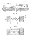
- Fig. 1 shows a vertical section through the diagnostic device of this invention; and
- Figs. 2 and 3 are sectional views through the device on line 2-2 to show different configurations of the containing means.
- Although the device and method of this invention are broadly applicable to determining the concentration of a select antigen or hapten in a test fluid, whereby said antigen also encompasses antibody protein, said antigen or hapten having the property of interacting specifically with an antibody, the invention will be described hereinbelow in the measurement of the level of immunoglobulin IgG in human serum. The apparatus is exemplary and, for example, the diagnostic substrate need not be planar in configurations, but may be stepped or curved.
- A generally channel-
shaped holder 10 having spaced impermeableopposed walls 11, 12 andbottom 13 having a flat upper surface serves to retain the test liquid. - End walls are also provided.
Ledge 14 is formed along wall 11 to provide support for the lower end of inclined diagnostic substrate 16 (a glass slide having indium layer 16a thereon with adsorbed layer 16b of purified human IgG thereover) shown with the upper end thereof resting on the top ofwall 12. The degree of inclination is exaggerated for ease of illustration. In the position shown a generally wedge-shaped volume is defined between slide (or diagnostic substrate) 16,wall 12,bottom 13 andvertical surface 17. - The containing
means 18 is made of a suitable liquid-absorbing material (i.e., porous and nonreactive), such as cellulose, acetate. Preferably the containing means (or moisture holding medium) is affixed to the upper surface ofbottom 13 by means of transferadhesive tape 19 and, when viewed in plan (Figs. 2 and 3), is formed in a convoluted or serpentine configuration affixed flat against the planar surface ofbottom 13. The containing means may, for example, be readily cut or stamped from a thin sheet of cellulose acetate or cellulose. Also, the containing means may comprise a gel disposed in a recess inbottom 13. -
Slide 16, when in place with its ends resting onledge 14 andwall 12, is disposed at a slight angle to the plane of containingmeans 18, this angle preferably being in the range of from about 3 minutes to about 10 degrees. Although this angle providing the wedge-like definition of volume is relatively small, it is sufficient (when the test liquid, a dilution of human serum, 21 is in place) to provide significant differentiation between the extent of arrival of anti-human IgG molecules at the lower end of layer 16b as contrasted to the higher end thereof. - It is preferred that the phenomenon of transport of the anti-human IgG molecules from absorbing
means 18 through the superimposedhuman serum 21 be by substantially pure diffusion and for this reason it may be necessary to minimize convection in the superimposed liquid. This can be done by assuring sufficiently high viscosity of the test liquid. If convection is properly reduced to a negligible amount with respect to the rate of diffusion, the reproduction of IgG layer 16b of the repeating pattern in which the absorbingmeans 18 has been shaped will occur as a second layer (not shown) having distinct lines. If the lines delineating the reproduction are fuzzy, a first approach is to increase the viscosity of the human serum. With aqueous solutions, appropriate increases in viscosity may be effectuated by introducing the liquid into a hydroxypropyl methyl cellulose solution or a glycerol solution. If this does not cure the problem of fuzziness, a less dilute sample is employed. - Although each of Figs. 2 and 3 shows a single strip in place of containing means 18 cut in a repeating pattern, the number of strips may be increased as desired with each of the
strips 18 receiving a different concentration of anti-human IgG serum for absorption therein. In a variant thestrips 18 may receive different anit-human serums, the slide being provided with a separate strip of antigen or hapten relating to the particular strip below it. By this means simultaneous tests may be conducted for IgG, IgM and IgA, for example. - In the method of this invention, a one-step inhibition test as presently conducted, a strip of cellulose acetate paper in some desired shape is first attached with
transfer tape 19 to the flat surface of bottom 13 as shown. Anti-human IgG serum is applied topaper 18 and is absorbed therein. Following absorption, excess anti-human IgG serum is blotted away. The human serum whose IgG level is to be measured is diluted in a 1% hydroxypropyl methyl cellulose solution and a small volume of the mixture (e.g., approximately 0.2 ml) is pipetted directly on the cellulose acetate paper. Finally, slide 16 consisting of a flat piece of glass, layer 16a of indium thereon and layer 16b of purified human IgG adsorbed on indium layer 16a is placed with opposite ends thereof onledge 14 andwall 12 and with layer 16b facing down as shown. The disposition ofslide 16 in this manner repositions thehuman serum dilution 21 into a wedge-shaped zone, the amount of this liquid previously deposited being as much or slightly more than will fill the volume betweenslide 16 and the walls ofdevice 10. - As the anti-human IgG serum diffuses from the cellulose acetate up into this liquid wedge, the specific antibodies comprising the anti-human serum complex with human IgG molecules (to the extent they are present in the test liquid 21) and are unable to proceed further toward
slide 16. In time, depending on the concentration of IgG in the liquid wedge, enough free antibodies (second biological particles) are able to diffuse upwardly to IgG layer 16b where they will complex with the molecules of layer 16b and form a second, visible layer thereon. This event first occurs at the thin edge of the liquid wedge (i.e., near the lower end of slide 16) where there is less human IgG disposed between thecellulose acetate paper 18 and layer 16b ofslide 16. As more time is allowed for diffusion to occur, the complexing event will gradually occur at thicker regions of the liquid wedge. After removing the slide, it is washed and dried (as by blowing). - Determining the extent (i.e., length and/or area) of the second layer formed on the diagnostic substrate is facilitated, if the
cellulose acetate strip 18 is cut in a repeating pattern as, for example, is shown in Figs. 2 and 3. - It has been found that with the construction shown in Figs. 1 and 2 in the drawing in which
ledge 14 andwall 12 are spaced one inch apart and the heights ofledge 14 and the top ofwall 12 from the upper surface of bottom 13 are 0.25 mm and 0.50 mm respectively, when using a 1:100 dilution of control human serum in a 1% hydroxypropyl methyl cellulose solution, a layer of purified IgG molecules on a coating of indium, undiluted anti-human IgG (whole molecule) serum from a rabbit and a 20 minute diffusion period, the anti-human IgG layer is visible to approximately half the length ofslide 16. The test conducted in this manner should reproducibly measure IgG concentrations of .04-.4 mg/ml with acceptable accuracy. - If, upon conducting the test, no second layer is manifest (indicating a very high level of IgG in the patient's serum), then the test could be repeated employing a greater dilution of the liquid sample to obtain some quantitative value. If, on the contrary, forthe time allotted for diffusion, the full pattern of the cellulose acetate paper is reproduced as a second layer (indicating a very low level of IgG in the patient's serum), then it will be necessary to repeat the test employing a more dilute solution of the antihuman IgG serum to obtain a quantitative value.
- The concentration of the select antigen or hapten solution used is so selected that the range of interest of the concentration of select antigen or hapten will register on the slide. Usually to conduct the test a first run is made with control serum (e.g., 12 mg of IgG/ml of serum) to define what is normal and the normal range is set therefrom so that subsequent readings can be related thereto. The slide can be marked off in units for convenience in setting the range.
- Variations of the device disclosed are readily contemplated as, for example, hinging
slide 16 to eitherledge 14 orwall 12 or using a flat holder with separate supporting means for the diagnostic substrate. - Demonstrations of this immunological test have been demonstrated in the laboratory employing a device as shown in which opposing
ledge 14 andwall 12 are spaced approximately one inch apart and the heights ofledge 14 and the top ofwall 12 from the upper surface of bottom 13 are 0.25 mm and 0.50 mm respectively. An IgG-coated planar indium slide and a flat- bottomed holder with a cellulose acetate strip attached thereto were employed.
Claims (17)
removing said substrate;
Applications Claiming Priority (2)
| Application Number | Priority Date | Filing Date | Title |
|---|---|---|---|
| US06/023,695 US4181501A (en) | 1979-03-26 | 1979-03-26 | Method and apparatus for measuring antibody levels |
| US23695 | 1979-03-26 |
Publications (3)
| Publication Number | Publication Date |
|---|---|
| EP0025794A1 EP0025794A1 (en) | 1981-04-01 |
| EP0025794A4 EP0025794A4 (en) | 1981-08-28 |
| EP0025794B1 true EP0025794B1 (en) | 1985-07-17 |
Family
ID=21816688
Family Applications (1)
| Application Number | Title | Priority Date | Filing Date |
|---|---|---|---|
| EP80900626A Expired EP0025794B1 (en) | 1979-03-26 | 1980-10-08 | Method and apparatus for measuring antibody levels |
Country Status (5)
| Country | Link |
|---|---|
| US (1) | US4181501A (en) |
| EP (1) | EP0025794B1 (en) |
| JP (1) | JPS6339872B2 (en) |
| DE (1) | DE3070876D1 (en) |
| WO (1) | WO1980002077A1 (en) |
Families Citing this family (13)
| Publication number | Priority date | Publication date | Assignee | Title |
|---|---|---|---|---|
| SE427389B (en) * | 1981-03-02 | 1983-03-28 | Alfa Laval Ab | INDICATOR INCLUDING A CAREER AND A REACTION SYSTEM |
| US4446232A (en) * | 1981-10-13 | 1984-05-01 | Liotta Lance A | Enzyme immunoassay with two-zoned device having bound antigens |
| DE3215484A1 (en) * | 1982-04-26 | 1983-11-03 | Sagax Instrument AB, 18302 Täby | MULTIPLE LAYERS OF LAYER AND PROCESS FOR DETECTING AND / OR MEASURING THE CONCENTRATION OF A CHEMICAL SUBSTANCE, IN PARTICULAR BIOLOGICAL ORIGIN |
| GB8629740D0 (en) * | 1986-12-12 | 1987-01-21 | Iq Bio Ltd | Immunoassay |
| FR2621393B1 (en) * | 1987-10-05 | 1991-12-13 | Toledano Jacques | DEVICE FOR IMMUNOENZYMATIC DETECTION OF SUBSTANCES FROM A DROP OF BLOOD OR LIQUID FROM ANY BIOLOGICAL MEDIUM |
| JPH01126474U (en) * | 1988-02-22 | 1989-08-29 | ||
| US5089387A (en) * | 1988-07-07 | 1992-02-18 | Adeza Biomedical Corporation | Dna probe diffraction assay and reagents |
| AU635008B2 (en) * | 1989-12-13 | 1993-03-11 | Genelabs Diagnostics Pte Ltd | Analytical apparatus and method for automated blot assay |
| US5192503A (en) * | 1990-05-23 | 1993-03-09 | Mcgrath Charles M | Probe clip in situ assay apparatus |
| US5413939A (en) * | 1993-06-29 | 1995-05-09 | First Medical, Inc. | Solid-phase binding assay system for interferometrically measuring analytes bound to an active receptor |
| US5679579A (en) * | 1996-01-29 | 1997-10-21 | First Medical, Inc. | Immunofluorescence measurement of analytes bound to a substrate and apparatus therefor |
| US5970782A (en) * | 1997-05-16 | 1999-10-26 | Life Technologies, Inc. | Gradient filtration apparatus |
| DK2988311T3 (en) | 2014-08-22 | 2021-07-26 | Abb Schweiz Ag | Pressure compensated subsea electrical system |
Citations (6)
| Publication number | Priority date | Publication date | Assignee | Title |
|---|---|---|---|---|
| US3770380A (en) * | 1971-04-19 | 1973-11-06 | Us Army | Article and method for multiple immune adherence assay |
| US3933594A (en) * | 1974-08-16 | 1976-01-20 | Polaroid Corporation | Method and device for determining the concentration of a substance in a fluid |
| US4018662A (en) * | 1975-01-03 | 1977-04-19 | Max-Planck-Gesellschaft Zur Forderung Der Wissenschaften E.V. | Method and apparatus for simultaneous quantitative analysis of several constituents in a sample |
| US4067959A (en) * | 1976-05-10 | 1978-01-10 | International Diagnostic Technology, Inc. | Indirect solid surface test for antigens or antibodies |
| FR2378283A1 (en) * | 1977-01-21 | 1978-08-18 | Searle & Co | Reaction testing mould - has base plate provided with depending plasma test tubes with support feet carrying removable identification cards (BE 24.7.78) |
| US4147752A (en) * | 1977-01-14 | 1979-04-03 | Kommandiittihytio Finnpipette Osmo A. Souvaniemi | Form piece for apparatuses used for immunoassays and enzyme reactions |
Family Cites Families (13)
| Publication number | Priority date | Publication date | Assignee | Title |
|---|---|---|---|---|
| US3674438A (en) * | 1970-05-20 | 1972-07-04 | James T Shen | Ouchterlony technique apparatus |
| US3672845A (en) * | 1970-07-28 | 1972-06-27 | Miles Lab | Test device for albumin |
| US3993451A (en) * | 1970-07-28 | 1976-11-23 | Miles Laboratories, Inc. | Test for a given constituent in a liquid |
| US3692486A (en) * | 1971-07-12 | 1972-09-19 | Cybertek Inc | Methods and apparatus for obtaining the quantitation and the concentrations of precipitin reactions and participating molecules in biological fluids |
| US3856628A (en) * | 1972-05-17 | 1974-12-24 | Crowley R | Method and apparatus for the identification of microorganisms |
| US4054646A (en) * | 1973-07-30 | 1977-10-18 | General Electric | Method and apparatus for detection of antibodies and antigens |
| ZA733612B (en) * | 1972-10-11 | 1974-04-24 | Merck Patent Gmbh | Container for test strips |
| US3853467A (en) * | 1973-08-15 | 1974-12-10 | Gen Electric | Method and apparatus for immunological detection of biological particles |
| US3926564A (en) * | 1974-02-25 | 1975-12-16 | Gen Electric | Substrate for immunological tests and method of fabrication thereof |
| SE403382B (en) * | 1974-03-12 | 1978-08-14 | Orion Yhtyme Oy Orion Diagnost | INVESTIGATE THE EFFECT OF A BIOLOGICAL ACTIVE SUBJECT ON THE GROWTH OF MICRO-ORGANISMS CULTIVATED ON A SOLID OR YELLOW-CULTURED MEDIUM |
| US3960490A (en) * | 1974-04-01 | 1976-06-01 | General Electric Company | Method and apparatus for detecting immunologic reactions by diffusion in gel |
| DE2416047A1 (en) * | 1974-04-03 | 1975-10-09 | Ludwig Clarius Wolfgang Dipl P | Transparent test strips for analysis of solutions - have varying thickness of gel giving visually evaluated test result |
| US3979184A (en) * | 1975-05-27 | 1976-09-07 | General Electric Company | Diagnostic device for visually detecting presence of biological particles |
-
1979
- 1979-03-26 US US06/023,695 patent/US4181501A/en not_active Expired - Lifetime
-
1980
- 1980-03-17 DE DE8080900626T patent/DE3070876D1/en not_active Expired
- 1980-03-17 WO PCT/US1980/000275 patent/WO1980002077A1/en active IP Right Grant
- 1980-03-17 JP JP55500751A patent/JPS6339872B2/ja not_active Expired
- 1980-10-08 EP EP80900626A patent/EP0025794B1/en not_active Expired
Patent Citations (6)
| Publication number | Priority date | Publication date | Assignee | Title |
|---|---|---|---|---|
| US3770380A (en) * | 1971-04-19 | 1973-11-06 | Us Army | Article and method for multiple immune adherence assay |
| US3933594A (en) * | 1974-08-16 | 1976-01-20 | Polaroid Corporation | Method and device for determining the concentration of a substance in a fluid |
| US4018662A (en) * | 1975-01-03 | 1977-04-19 | Max-Planck-Gesellschaft Zur Forderung Der Wissenschaften E.V. | Method and apparatus for simultaneous quantitative analysis of several constituents in a sample |
| US4067959A (en) * | 1976-05-10 | 1978-01-10 | International Diagnostic Technology, Inc. | Indirect solid surface test for antigens or antibodies |
| US4147752A (en) * | 1977-01-14 | 1979-04-03 | Kommandiittihytio Finnpipette Osmo A. Souvaniemi | Form piece for apparatuses used for immunoassays and enzyme reactions |
| FR2378283A1 (en) * | 1977-01-21 | 1978-08-18 | Searle & Co | Reaction testing mould - has base plate provided with depending plasma test tubes with support feet carrying removable identification cards (BE 24.7.78) |
Also Published As
| Publication number | Publication date |
|---|---|
| JPS6339872B2 (en) | 1988-08-08 |
| EP0025794A1 (en) | 1981-04-01 |
| EP0025794A4 (en) | 1981-08-28 |
| US4181501A (en) | 1980-01-01 |
| WO1980002077A1 (en) | 1980-10-02 |
| DE3070876D1 (en) | 1985-08-22 |
| JPS56500310A (en) | 1981-03-12 |
Similar Documents
| Publication | Publication Date | Title |
|---|---|---|
| US5569608A (en) | Quantitative detection of analytes on immunochromatographic strips | |
| EP1075661B1 (en) | Ligand binding assay and kit with a separation zone for disturbing sample components | |
| JP2948318B2 (en) | Red blood cell separation method for specific binding assays | |
| US5416000A (en) | Analyte immunoassay in self-contained apparatus | |
| EP0335244B1 (en) | Solid-phase analytical device and method for using same | |
| US5492840A (en) | Surface plasmon resonance sensor unit and its use in biosensor systems | |
| EP1634078B1 (en) | Native analyte as reference in lateral flow assays | |
| US7521196B2 (en) | Prewetting lateral flow test strip | |
| EP0025794B1 (en) | Method and apparatus for measuring antibody levels | |
| AU610818B2 (en) | Blood typing via immunological device | |
| JPH0627738B2 (en) | Specific binding assay device and method | |
| JP4328203B2 (en) | Multiple-analyte assay device in multiple-spot detection zone | |
| EP0207152B1 (en) | Solid phase diffusion assay | |
| EP0888547B1 (en) | Immunoassay with whole blood | |
| EP0972073A1 (en) | Diagnostic test devices with improved fluid movement and resistance to interferences | |
| JP2505658B2 (en) | Test carrier for analytical measurement of sample fluid components | |
| CA2198948A1 (en) | Quantitative detection of analytes on immunochromatographic strips | |
| Glad et al. | A new method for immunochemical quantification | |
| JPS5943360A (en) | Immunological measuring method | |
| JPS60127462A (en) | Enzymatic immune measuring method | |
| JP2004108783A (en) | Solid analysis medium | |
| CA2149537A1 (en) | Method for preparing calibration curves | |
| JPS61213672A (en) | Immunoassay method |
Legal Events
| Date | Code | Title | Description |
|---|---|---|---|
| PUAI | Public reference made under article 153(3) epc to a published international application that has entered the european phase |
Free format text: ORIGINAL CODE: 0009012 |
|
| 17P | Request for examination filed |
Effective date: 19810109 |
|
| AK | Designated contracting states |
Designated state(s): DE FR GB |
|
| GRAA | (expected) grant |
Free format text: ORIGINAL CODE: 0009210 |
|
| AK | Designated contracting states |
Designated state(s): DE FR GB |
|
| REF | Corresponds to: |
Ref document number: 3070876 Country of ref document: DE Date of ref document: 19850822 |
|
| ET | Fr: translation filed | ||
| PLBE | No opposition filed within time limit |
Free format text: ORIGINAL CODE: 0009261 |
|
| STAA | Information on the status of an ep patent application or granted ep patent |
Free format text: STATUS: NO OPPOSITION FILED WITHIN TIME LIMIT |
|
| 26N | No opposition filed | ||
| PGFP | Annual fee paid to national office [announced via postgrant information from national office to epo] |
Ref country code: GB Payment date: 19901219 Year of fee payment: 12 |
|
| PGFP | Annual fee paid to national office [announced via postgrant information from national office to epo] |
Ref country code: FR Payment date: 19901228 Year of fee payment: 12 |
|
| PGFP | Annual fee paid to national office [announced via postgrant information from national office to epo] |
Ref country code: DE Payment date: 19910403 Year of fee payment: 12 |
|
| PG25 | Lapsed in a contracting state [announced via postgrant information from national office to epo] |
Ref country code: GB Effective date: 19920317 |
|
| GBPC | Gb: european patent ceased through non-payment of renewal fee | ||
| PG25 | Lapsed in a contracting state [announced via postgrant information from national office to epo] |
Ref country code: FR Effective date: 19921130 |
|
| PG25 | Lapsed in a contracting state [announced via postgrant information from national office to epo] |
Ref country code: DE Effective date: 19921201 |
|
| REG | Reference to a national code |
Ref country code: FR Ref legal event code: ST |